

Co-infection of classic swine H1N1 influenza virus in pigs persistently infected with porcine rubulavirus
- PMID: 26854342
- PMCID: PMC7117528
- DOI: 10.1016/j.vetmic.2016.01.005
Co-infection of classic swine H1N1 influenza virus in pigs persistently infected with porcine rubulavirus
Abstract
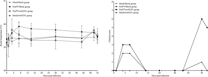
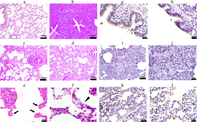
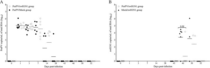
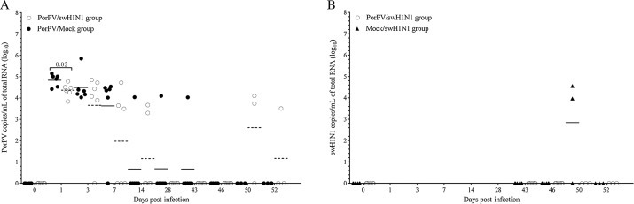
Porcine rubulavirus (PorPV) and swine influenza virus infection causes respiratory disease in pigs. PorPV persistent infection could facilitate the establishment of secondary infections. The aim of this study was to analyse the pathogenicity of classic swine H1N1 influenza virus (swH1N1) in growing pigs persistently infected with porcine rubulavirus. Conventional six-week-old pigs were intranasally inoculated with PorPV, swH1N1, or PorPV/swH1N1. A mock-infected group was included. The co-infection with swH1N1 was at 44 days post-infection (DPI), right after clinical signs of PorPV infection had stopped. The pigs of the co-infection group presented an increase of clinical signs compared to the simple infection groups. In all infected groups, the most recurrent lung lesion was hyperplasia of the bronchiolar-associated lymphoid tissue and interstitial pneumonia. By means of immunohistochemical evaluation it was possible to demonstrate the presence of the two viral agents infecting simultaneously the bronchiolar epithelium. Viral excretion of PorPV in nasal and oral fluid was recorded at 28 and 52 DPI, respectively. PorPV persisted in several samples from respiratory tissues (RT), secondary lymphoid organs (SLO), and bronchoalveolar lavage fluid (BALF). For swH1N1, the viral excretion in nasal fluids was significantly higher in single-infected swH1N1 pigs than in the co-infected group. However, the co-infection group exhibited an increase in the presence of swH1N1 in RT, SLO, and BALF at two days after co-infection. In conclusion, the results obtained confirm an increase in the clinical signs of infection, and PorPV was observed to impact the spread of swH1N1 in analysed tissues in the early stage of co-infection, although viral shedding was not enhanced. In the present study, the interaction of swH1N1 infection is demonstrated in pigs persistently infected with PorPV.
Keywords: Classic swine H1N1 influenza virus; Co-infection; Pigs; Porcine rubulavirus; Respiratory disease.
Copyright © 2016 Elsevier B.V. All rights reserved.
Figures
Similar articles
-
Respiratory disease in growing pigs after Porcine rubulavirus experimental infection.Virus Res. 2013 Sep;176(1-2):137-43. doi: 10.1016/j.virusres.2013.05.017. Epub 2013 Jun 12. Virus Res. 2013. PMID: 23770154
-
Long-term RNA persistence of porcine rubulavirus (PorPV-LPMV) after an outbreak of a natural infection: the detection of viral mRNA in sentinel pigs suggests viral transmission.Virus Res. 2014 Aug 8;188:155-61. doi: 10.1016/j.virusres.2014.04.012. Epub 2014 Apr 24. Virus Res. 2014. PMID: 24768705
-
Persistence of porcine rubulavirus in experimentally infected boars.Vet Microbiol. 2013 Mar 23;162(2-4):491-498. doi: 10.1016/j.vetmic.2012.10.037. Epub 2012 Nov 11. Vet Microbiol. 2013. PMID: 23201243
-
Acute neurologic disease in Porcine rubulavirus experimentally infected piglets.Virus Res. 2017 Feb 15;230:50-58. doi: 10.1016/j.virusres.2017.01.010. Epub 2017 Jan 16. Virus Res. 2017. PMID: 28104449
-
Molecular characterisation of Porcine rubulavirus (PorPV) isolates from different outbreaks in Mexico.Virus Genes. 2016 Feb;52(1):81-90. doi: 10.1007/s11262-015-1281-y. Epub 2016 Jan 4. Virus Genes. 2016. PMID: 26728078
Cited by
-
Development of Novel Recombinant Antigens of Nucleoprotein and Matrix Proteins of Porcine orthorubulavirus: Antigenicity and Structural Prediction.Viruses. 2022 Sep 1;14(9):1946. doi: 10.3390/v14091946. Viruses. 2022. PMID: 36146753 Free PMC article.
References
-
- Avalos G., Sanchez B., Trujillo O. 1st edition. Editorial Académica Española; Saarbrücken, Germany: 2011. Influenza Porcina en México; p. 80.
-
- Bobadilla S.E., Espinoza O.A., Martínez C.F. Dinámica de la producción porcina en México de 1980 a 2008. Revista Mexicana de Ciencias Pecuarias. 2010;1:251–268.
-
- Brookes S.M., Irvine R.M., Nunez A., Clifford D., Essen S., Brown I.H., Van Reeth K., Kuntz-Simon G., Loeffen W., Foni E., Larsen L., Matrosovich M., Bublot M., Maldonado J., Beer M., Cattoli G. Influenza A (H1N1) infection in pigs. Vet. Rec. 2009;164:760–761. - PubMed
-
- Busquets N., Segales J., Cordoba L., Mussa T., Crisci E., Martin-Valls G.E., Simon-Grife M., Perez-Simo M., Perez-Maillo M., Nunez J.I., Abad F.X., Fraile L., Pina S., Majo N., Bensaid A., Domingo M., Montoya M. Experimental infection with H1N1 European swine influenza virus protects pigs from an infection with the 2009 pandemic H1N1 human influenza virus. Vet. Res. 2010;41:74. - PMC - PubMed
Publication types
MeSH terms
Substances
LinkOut - more resources
Full Text Sources
Other Literature Sources
