. 2016 Feb 5:5:e12129.
doi: 10.7554/eLife.12129.

Preservation of three-dimensional anatomy in phosphatized fossil arthropods enriches evolutionary inference

Affiliations

Preservation of three-dimensional anatomy in phosphatized fossil arthropods enriches evolutionary inference

Achim H Schwermann et al. Elife. .

Abstract

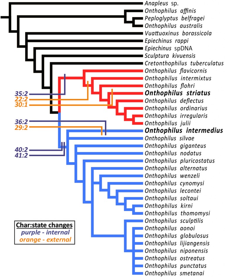
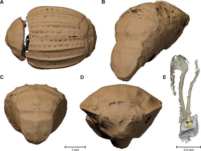
External and internal morphological characters of extant and fossil organisms are crucial to establishing their systematic position, ecological role and evolutionary trends. The lack of internal characters and soft-tissue preservation in many arthropod fossils, however, impedes comprehensive phylogenetic analyses and species descriptions according to taxonomic standards for Recent organisms. We found well-preserved three-dimensional anatomy in mineralized arthropods from Paleogene fissure fillings and demonstrate the value of these fossils by utilizing digitally reconstructed anatomical structure of a hister beetle. The new anatomical data facilitate a refinement of the species diagnosis and allowed us to reject a previous hypothesis of close phylogenetic relationship to an extant congeneric species. Our findings suggest that mineralized fossils, even those of macroscopically poor preservation, constitute a rich but yet largely unexploited source of anatomical data for fossil arthropods.

Keywords: Histeridae; Paleogene; X-ray imaging; evolutionary biology; fissure fillings; genomics; internal characters; onthophilus spp. (coleopera: histeridae).


Conflict of interest statement

The authors declare that no competing interests exist.

Figures

Figure 1.
Figure 1.. Comparison between the fossil Onthophilus intermedius (ADG) and EtOH-fixed (BEH) and air-dried (CF , I) specimens of O. striatus.
Slices of tomographic volumes showing head region (AC), thorax (D–F) and abdomen (G–I). ae = aedeagus; ag = accessory gland; bpae = basal part of aedeagus; hg = hindgut; m = musculature; ml = median lobe; mr = muscles remnants; mscx = mesocoxa; msf = mesofemur; mst = mesotibia; mt = muscle tissue; mtcx = metacoxa; mtf = metafemur; mtt = metatibia; pcx = procoxa; sph = spherical particle; sm = stony matrix; t8 = 8th abdominal tergite; t9 = 9th abdominal tergite; t10 = 10th abdominal tergite; te = tentorium; tr = trachea. DOI: http://dx.doi.org/10.7554/eLife.12129.003
Figure 2.
Figure 2.. Surface renderings of the eight Onthophilus intermedius specimens.
Note the unique encrustation of F1994. DOI: http://dx.doi.org/10.7554/eLife.12129.005
Figure 3.
Figure 3.. Digital reconstruction of the fossil.
(A) Photograph of Onthophilus intermedius (F1994) ventrally embedded in a stony matrix. (B) Digital reconstruction showing fossilized beetle (green) and matrix (brown). (C) Beetle digitally isolated from the stone, revealing well-preserved morphology hidden by the matrix. (D) Perspective view of the fossil showing parts of exoskeleton, tracheal network, alimentary canal and genitals. (E, F) Comparison of the male genitals of the extant O. striatus (E) and the fossil O. intermedius (F); outer sclerites cut to reveal internal anatomy. See Supplementary file 1 for an interactive version of the 3D reconstruction. DOI: http://dx.doi.org/10.7554/eLife.12129.006
Figure 4.
Figure 4.. Coxa-trochanteral joints.
Comparison of the joints (cut) of the left mid- (AB) and hind leg (CD) of Onthophilus striatus (AC) and O. intermedius (BD), showing coxae (green) and trochanters (yellow). DOI: http://dx.doi.org/10.7554/eLife.12129.007
Figure 5.
Figure 5.. Digital endocast of Onthophilus intermedius (specimen F1994).
A digital endocast (AB) artificially created from tomography data resembles the shape of the other fossils (Figure 2) much closer than the original surface of the beetle (CD) hidden by the stony matrix. DOI: http://dx.doi.org/10.7554/eLife.12129.008
Figure 6.
Figure 6.. Strict consensus tree.
The analysis places Onthophilus striatus within a lineage of Nearctic and far-eastern Palaearctic species (red), while O. intermedius is a member of a separate Holarctic lineage (blue). Four internal (purple) and three external (orange) unambiguous synapomorphies supporting their respective placements are mapped onto the cladogram - Onthophilus striatus group: Character 22:2, mesoventrite wide and short; 30:1, pygidial median carina absent; 35:2, tegmen of aedeagus abruptly downturned apically. O. intermedius group: 29:2, pygidium laterally impunctate; 36:2, tegmen of aedeagus abruptly narrowing apically; 40:2, lateral halves of eighth sternite large and nearly meeting at midline; 41:2, stem of spiculum gastrale broad throughout its length. DOI: http://dx.doi.org/10.7554/eLife.12129.009
Appendix figure 1.
Appendix figure 1.. Morphological characters visible in specimens other than F1994.
(A) Dorsal view of F1951, showing elytral carinae and foveae. (B) Posterolateral view of F1997, showing propygidial and pygidial carinae. (C) Posterior view of F1951. (D) Anterior view of F1995. (E) Valvifer and coxite of female ovipositor. DOI: http://dx.doi.org/10.7554/eLife.12129.012

References

    1. Allison PA, Bottjer DJ. Taphonomy: bias and process through time. In: Allison PA, Bottjer DJ, editors. Taphonomy. Dordrecht: Springer Netherlands; 2011. pp. 1–17. - DOI
    1. Anderson LI, Trewin NH. An Early Devonian arthropod fauna from the Windyfield cherts, Aberdeenshire, Scotland. Palaeontology. 2003;46:467–509. doi: 10.1111/1475-4983.00308. - DOI
    1. Bajerlein D, Matuszewski S, Konwerski S. Insect succession on carrion: seasonality, habitat preference and residency of histerid beetles (Coleoptera: Histeridae) visiting pig carrion exposed in various forests (Western Poland) Polish Journal of Ecology. 2011;59:787–797.
    1. Barling N, Martill DM, Heads SW, Gallien F. High fidelity preservation of fossil insects from the Crato Formation (Lower Cretaceous) of Brazil. Cretaceous Research. 2015;52:605–622. doi: 10.1016/j.cretres.2014.05.007. - DOI
    1. Betz O, Wegst U, Weide D, Heethoff M, Helfen L, Lee W-K, Cloetens P. Imaging applications of synchrotron X-ray phase-contrast microtomography in biological morphology and biomaterials science. I. General aspects of the technique and its advantages in the analysis of millimetre-sized arthropod structure. Journal of Microscopy. 2007;227:51–71. doi: 10.1111/j.1365-2818.2007.01785.x. - DOI - PubMed