

The effect of local anesthetic on pro-inflammatory macrophage modulation by mesenchymal stromal cells
- PMID: 26854576
- PMCID: PMC4779686
- DOI: 10.1016/j.intimp.2016.01.019
The effect of local anesthetic on pro-inflammatory macrophage modulation by mesenchymal stromal cells
Abstract
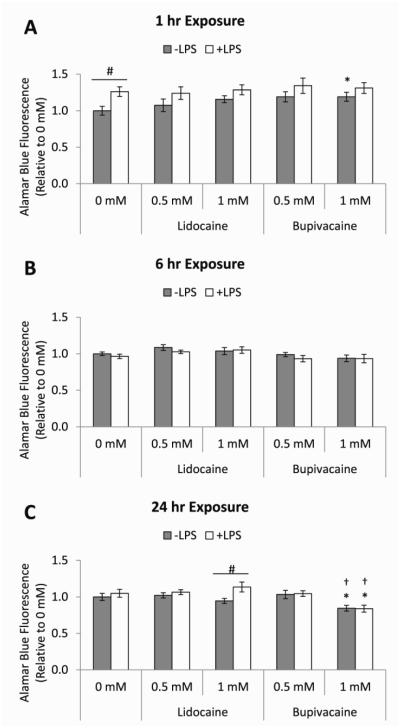
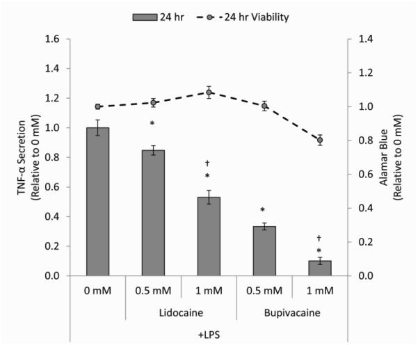
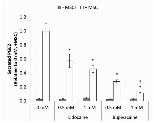
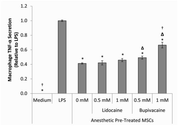
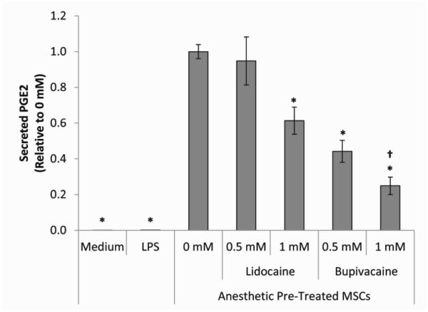
Administering local anesthetics (LAs) peri- and post-operatively aims to prevent or mitigate pain in surgical procedures and after tissue injury in cases of osteoarthritis (OA) and other degenerative diseases. Innovative tissue protective and reparative therapeutic interventions such as mesenchymal stromal cells (MSCs) are likely to be exposed to co-administered drugs such as LAs. Therefore, it is important to determine how this exposure affects the therapeutic functions of MSCs and other cells in their target microenvironment. In these studies, we measured the effect of LAs, lidocaine and bupivacaine, on macrophage viability and pro-inflammatory secretion. We also examined their effect on modulation of the macrophage pro-inflammatory phenotype in an in vitro co-culture system with MSCs, by quantifying macrophage tumor necrosis factor (TNF)-α secretion and MSC prostaglandin E2 (PGE2) production. Our studies indicate that both LAs directly attenuated macrophage TNF-α secretion, without significantly affecting viability, in a concentration- and potency-dependent manner. LA-mediated attenuation of macrophage TNF-α was sustained in co-culture with MSCs, but MSCs did not further enhance this anti-inflammatory effect. Concentration- and potency-dependent reductions in macrophage TNF-α were concurrent with decreased PGE2 levels in the co-cultures further indicating MSC-independent macrophage attenuation. MSC functional recovery from LA exposure was assessed by pre-treating MSCs with LAs prior to co-culture with macrophages. Both MSC attenuation of TNF-α and PGE2 secretion were impaired by pre-exposure to the more potent bupivacaine and high dose of lidocaine in a concentration-dependent manner. Therefore, LAs can affect anti-inflammatory function by both directly attenuating macrophage inflammation and MSC secretion and possibly by altering the local microenvironment which can secondarily reduce MSC function. Furthermore, the LA effect on MSC function may persist even after LA removal.
Keywords: Inflammation; Local anesthetics; Macrophages; Mesenchymal stromal cells.
Copyright © 2016 Elsevier B.V. All rights reserved.
Figures

Similar articles
-
Pre-conditioning mesenchymal stromal cell spheroids for immunomodulatory paracrine factor secretion.Cytotherapy. 2014 Mar;16(3):331-45. doi: 10.1016/j.jcyt.2013.09.004. Epub 2013 Nov 9. Cytotherapy. 2014. PMID: 24219905
-
Identification of IL-1β and LPS as optimal activators of monolayer and alginate-encapsulated mesenchymal stromal cell immunomodulation using design of experiments and statistical methods.Biotechnol Prog. 2015 Jul-Aug;31(4):1058-70. doi: 10.1002/btpr.2103. Epub 2015 May 28. Biotechnol Prog. 2015. PMID: 25958832 Free PMC article.
-
Immunoregulatory potential of mesenchymal stem cells following activation by macrophage-derived soluble factors.Stem Cell Res Ther. 2019 Feb 13;10(1):58. doi: 10.1186/s13287-019-1156-6. Stem Cell Res Ther. 2019. PMID: 30760316 Free PMC article.
-
Mesenchymal Stem Cells Direct the Immunological Fate of Macrophages.Results Probl Cell Differ. 2017;62:61-72. doi: 10.1007/978-3-319-54090-0_4. Results Probl Cell Differ. 2017. PMID: 28455706 Review.
-
Mesenchymal stem cells maintain the microenvironment of central nervous system by regulating the polarization of macrophages/microglia after traumatic brain injury.Int J Neurosci. 2017 Dec;127(12):1124-1135. doi: 10.1080/00207454.2017.1325884. Epub 2017 May 19. Int J Neurosci. 2017. PMID: 28464695 Review.
Cited by
-
Perioperative Intravenous Lidocaine and Metastatic Cancer Recurrence - A Narrative Review.Front Oncol. 2021 Aug 2;11:688896. doi: 10.3389/fonc.2021.688896. eCollection 2021. Front Oncol. 2021. PMID: 34408981 Free PMC article. Review.
-
A Step-By-Step Surgical Protocol for the Treatment of Perianal Fistula with Adipose-Derived Mesenchymal Stem Cells.J Gastrointest Surg. 2018 Nov;22(11):2003-2012. doi: 10.1007/s11605-018-3895-6. Epub 2018 Jul 31. J Gastrointest Surg. 2018. PMID: 30066070
-
Cis-2-Decenoic Acid and Bupivacaine Delivered from Electrospun Chitosan Membranes Increase Cytokine Production in Dermal and Inflammatory Cell Lines.Pharmaceutics. 2023 Oct 17;15(10):2476. doi: 10.3390/pharmaceutics15102476. Pharmaceutics. 2023. PMID: 37896236 Free PMC article.
-
Lidocaine Potentiates SOCS3 to Attenuate Inflammation in Microglia and Suppress Neuropathic Pain.Cell Mol Neurobiol. 2019 Nov;39(8):1081-1092. doi: 10.1007/s10571-019-00703-6. Epub 2019 Jun 17. Cell Mol Neurobiol. 2019. PMID: 31209627 Free PMC article.
-
A Review of the Effects of Pain and Analgesia on Immune System Function and Inflammation: Relevance for Preclinical Studies.Comp Med. 2019 Dec 1;69(6):520-534. doi: 10.30802/AALAS-CM-19-000041. Epub 2019 Dec 20. Comp Med. 2019. PMID: 31896389 Free PMC article. Review.
References
-
- Breu A, et al. Cytotoxicity of local anesthetics on human mesenchymal stem cells in vitro. Arthroscopy. 2013;29(10):1676–84. - PubMed
-
- Rahnama R, et al. Cytotoxicity of local anesthetics on human mesenchymal stem cells. The Journal of bone and joint surgery. American volume. 2013;95(2):132–7. - PubMed
-
- Fahy N, et al. Human osteoarthritic synovium impacts chondrogenic differentiation of mesenchymal stem cells via macrophage polarisation state. Osteoarthritis and Cartilage. 2014;22(8):1167–75. - PubMed
Publication types
MeSH terms
Substances
Grants and funding
LinkOut - more resources
Full Text Sources
Other Literature Sources
Medical
