

Increased retinal mtDNA damage in the CFH variant associated with age-related macular degeneration
- PMID: 26854823
- PMCID: PMC4842097
- DOI: 10.1016/j.exer.2016.01.018
Increased retinal mtDNA damage in the CFH variant associated with age-related macular degeneration
Abstract
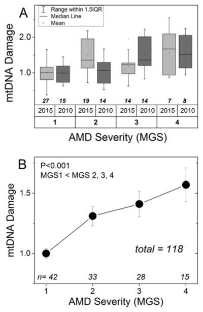
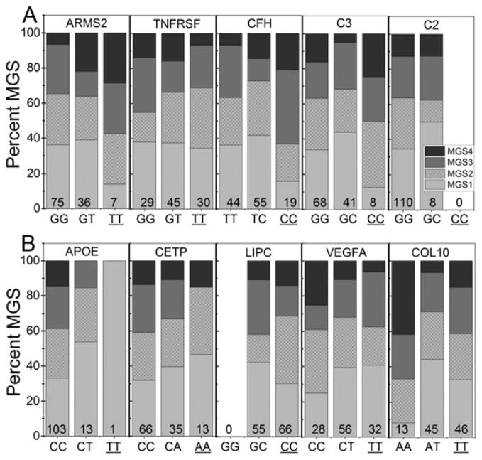
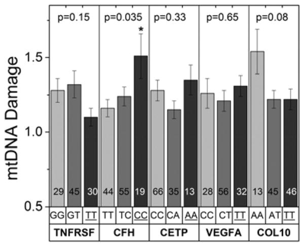
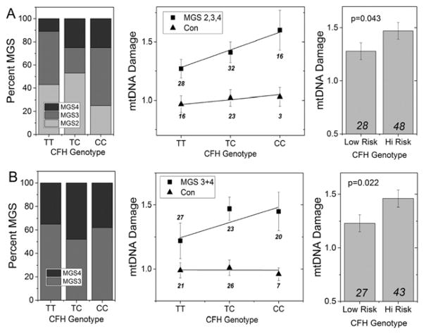
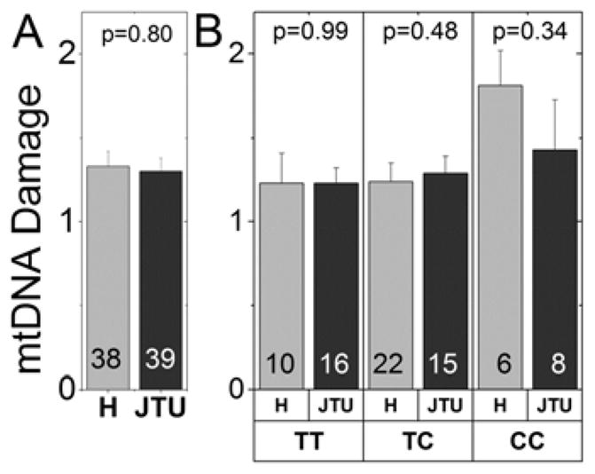
Age-related macular degeneration (AMD) is a major cause of blindness among the elderly in the developed world. Genetic analysis of AMD has identified 34 high-risk loci associated with AMD. The genes at these high risk loci belong to diverse biological pathways, suggesting different mechanisms leading to AMD pathogenesis. Thus, therapies targeting a single pathway for all AMD patients will likely not be universally effective. Recent evidence suggests defects in mitochondria (mt) of the retinal pigment epithelium (RPE) may constitute a key pathogenic event in some AMD patients. The purpose of this study is to determine if individuals with a specific genetic background have a greater propensity for mtDNA damage. We used human eyebank tissues from 76 donors with AMD and 42 age-matched controls to determine the extent of mtDNA damage in the RPE that was harvested from the macula using a long extension polymerase chain reaction assay. Genotype analyses were performed for ten common AMD-associated nuclear risk alleles (ARMS2, TNFRSF10A, CFH, C2, C3, APOE, CETP, LIPC, VEGF and COL10A1) and mtDNA haplogroups. Sufficient samples were available for genotype association with mtDNA damage for TNFRSF10A, CFH, CETP, VEGFA, and COL10A1. Our results show that AMD donors carrying the high risk allele for CFH (C) had significantly more mtDNA damage compared with donors having the wild-type genetic profile. The data from an additional 39 donors (12 controls and 27 AMD) genotyped for CFH alleles further supported these findings. Taken together, these studies provide the rationale for a more personalized approach for treating AMD by uncovering a significant correlation between the CFH high risk allele and accelerated mtDNA damage. Patients harboring this genetic risk factor may benefit from therapies that stabilize and protect the mt in the RPE.
Keywords: Age-related macular degeneration; Complement factor H; Eyebank tissue; Haplogroups; Inflammation; Mitochondria; mtDNA.
Copyright © 2016 Elsevier Ltd. All rights reserved.
Figures
Similar articles
-
Impaired Mitochondrial Function in iPSC-Retinal Pigment Epithelium with the Complement Factor H Polymorphism for Age-Related Macular Degeneration.Cells. 2021 Apr 2;10(4):789. doi: 10.3390/cells10040789. Cells. 2021. PMID: 33918210 Free PMC article.
-
The association between macular pigment optical density and CFH, ARMS2, C2/BF, and C3 genotype.Exp Eye Res. 2011 Nov;93(5):592-8. doi: 10.1016/j.exer.2011.07.005. Epub 2011 Jul 27. Exp Eye Res. 2011. PMID: 21816153
-
Risk alleles in CFH and ARMS2 are independently associated with systemic complement activation in age-related macular degeneration.Ophthalmology. 2012 Feb;119(2):339-46. doi: 10.1016/j.ophtha.2011.07.056. Epub 2011 Nov 30. Ophthalmology. 2012. PMID: 22133792
-
Hypothetical pathogenesis of age-related macular degeneration and pachychoroid diseases derived from their genetic characteristics.Jpn J Ophthalmol. 2020 Nov;64(6):555-567. doi: 10.1007/s10384-020-00773-w. Epub 2020 Oct 2. Jpn J Ophthalmol. 2020. PMID: 33006732 Review.
-
Age-related macular degeneration: Complement in action.Immunobiology. 2016 Jun;221(6):733-9. doi: 10.1016/j.imbio.2015.11.007. Epub 2015 Dec 19. Immunobiology. 2016. PMID: 26742632 Review.
Cited by
-
Retinal Manifestations of Mitochondrial Oxidative Phosphorylation Disorders.Invest Ophthalmol Vis Sci. 2020 Oct 1;61(12):12. doi: 10.1167/iovs.61.12.12. Invest Ophthalmol Vis Sci. 2020. PMID: 33049060 Free PMC article.
-
PU-91 drug rescues human age-related macular degeneration RPE cells; implications for AMD therapeutics.Aging (Albany NY). 2019 Sep 2;11(17):6691-6713. doi: 10.18632/aging.102179. Epub 2019 Sep 2. Aging (Albany NY). 2019. PMID: 31477635 Free PMC article.
-
Senescence in the pathogenesis of age-related macular degeneration.Cell Mol Life Sci. 2020 Mar;77(5):789-805. doi: 10.1007/s00018-019-03420-x. Epub 2020 Jan 2. Cell Mol Life Sci. 2020. PMID: 31897543 Free PMC article. Review.
-
Daily Light Onset and Plasma Membrane Tethers Regulate Mitochondria Redistribution within the Retinal Pigment Epithelium.Cells. 2024 Jun 25;13(13):1100. doi: 10.3390/cells13131100. Cells. 2024. PMID: 38994953 Free PMC article.
-
Impaired Mitochondrial Function in iPSC-Retinal Pigment Epithelium with the Complement Factor H Polymorphism for Age-Related Macular Degeneration.Cells. 2021 Apr 2;10(4):789. doi: 10.3390/cells10040789. Cells. 2021. PMID: 33918210 Free PMC article.
References
-
- Atilano SR, Malik D, Chwa M, Cáceres-Del-Carpio J, Nesburn AB, Boyer DS, Kuppermann BD, Jazwinski SM, Miceli MV, Wallace DC, et al. Mitochondrial DNA variants can mediate methylation status of inflammation, angiogenesis and signaling genes. Hum Mol Genet. 2015 doi: 10.1093/hmg/ddv173. - DOI - PMC - PubMed
-
- Chen W, Stambolian D, Edwards AO, Branham KE, Othman M, Jakobsdottir J, Tosakulwong N, Pericak-Vance MA, Campochiaro PA, Klein ML, et al. Genetic variants near TIMP3 and high-density lipoprotein-associated loci influence susceptibility to age-related macular degeneration. PNAS. 2010;107:7401–7406. - PMC - PubMed
-
- Chew EY, Klein ML, Clemons TE, Agrón E, Ratnapryiya R, Edwards AO, Fritsche LG, Swaroop A, Abecasis GR Age-Related Eye Disease Study Research Group. No clinically significant association between CFH and ARMS2 genotypes and response to nutritional supplements: AREDS report number 38. Ophthalmology. 2014;121:2173–2180. - PMC - PubMed
Publication types
MeSH terms
Substances
Grants and funding
LinkOut - more resources
Full Text Sources
Other Literature Sources
Medical
Miscellaneous
