. 2016 Mar 3;531(7592):114-117.
doi: 10.1038/nature16988. Epub 2016 Feb 8.

Cryo-electron microscopy structure of a coronavirus spike glycoprotein trimer

Affiliations

Cryo-electron microscopy structure of a coronavirus spike glycoprotein trimer

Alexandra C Walls et al. Nature. .

Abstract

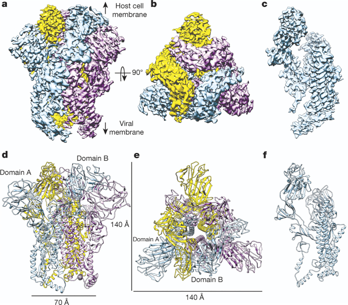
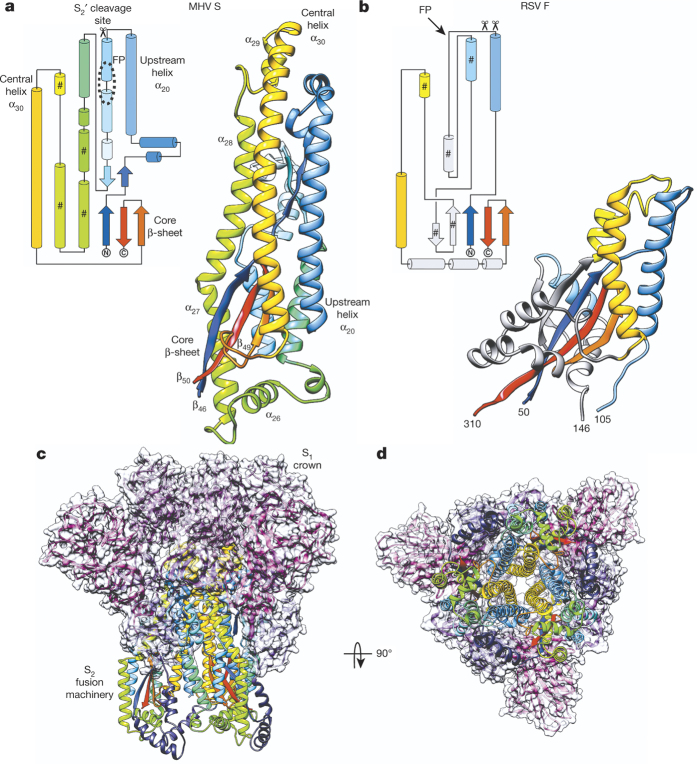
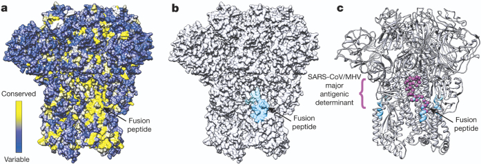
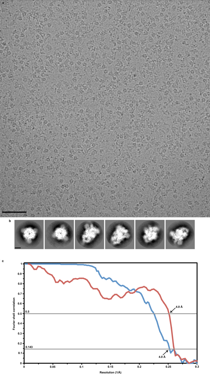
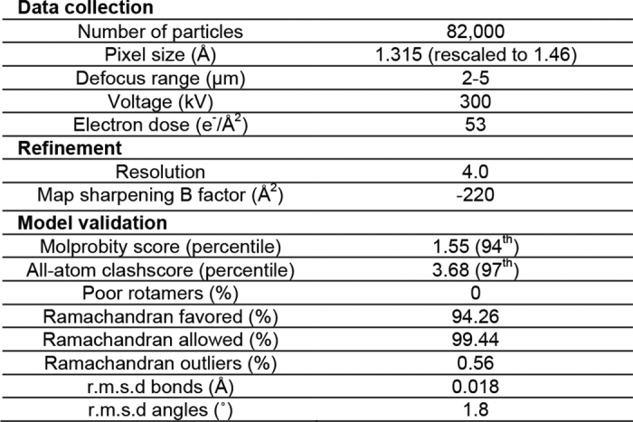
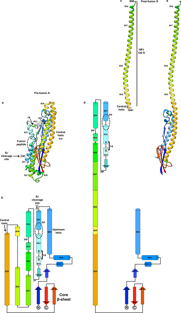
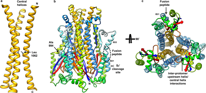
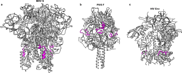
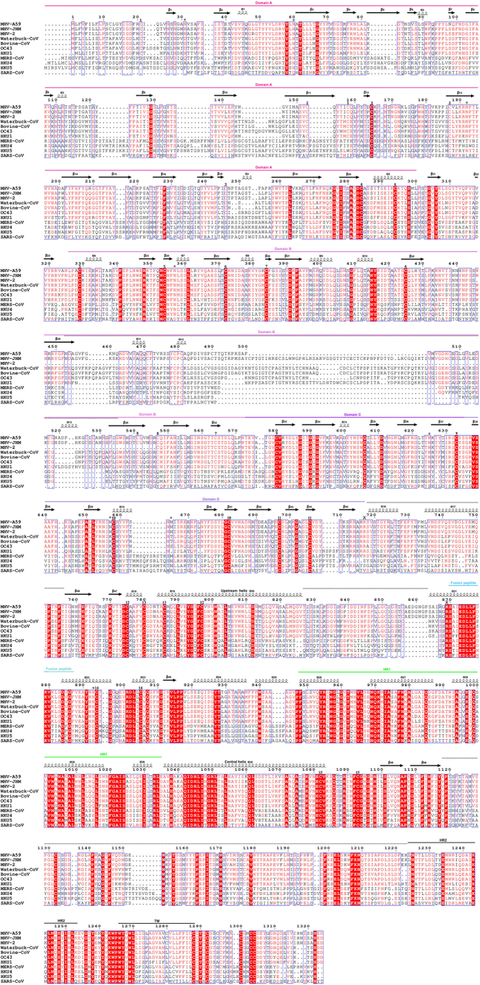
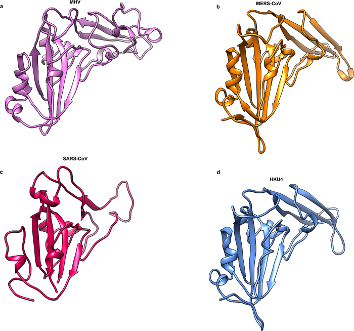
The tremendous pandemic potential of coronaviruses was demonstrated twice in the past few decades by two global outbreaks of deadly pneumonia. Entry of coronaviruses into cells is mediated by the transmembrane spike glycoprotein S, which forms a trimer carrying receptor-binding and membrane fusion functions. S also contains the principal antigenic determinants and is the target of neutralizing antibodies. Here we present the structure of a mouse coronavirus S trimer ectodomain determined at 4.0 Å resolution by single particle cryo-electron microscopy. It reveals the metastable pre-fusion architecture of S and highlights key interactions stabilizing it. The structure shares a common core with paramyxovirus F proteins, implicating mechanistic similarities and an evolutionary connection between these viral fusion proteins. The accessibility of the highly conserved fusion peptide at the periphery of the trimer indicates potential vaccinology strategies to elicit broadly neutralizing antibodies against coronaviruses. Finally, comparison with crystal structures of human coronavirus S domains allows rationalization of the molecular basis for species specificity based on the use of spatially contiguous but distinct domains.


Conflict of interest statement

The authors declare no competing financial interests.

Figures

Figure 1
Figure 1. 3D reconstruction of the MHV S trimer determined by single-particle cryoEM.
ac, 3D map filtered at 4.0 Å resolution coloured by protomer. Two different views of the S trimer (from the side (a) and from the top, looking towards the viral membrane (b)), and a side view of one S protomer (c) are shown. df, Ribbon diagrams showing the MHV S atomic model oriented as in ac. PowerPoint slide
Figure 2
Figure 2. Architecture of the MHV S protomer.
a, Schematic diagram of the S glycoprotein organization. Black and grey dashed lines denote regions unresolved in the reconstruction and regions that were not part of the construct, respectively. BH, β-hairpin (β49–β50); CH, central helix; CT, cytoplasmic tail; FP, fusion peptide; HR1/HR2, heptad-repeats; TM, transmembrane domain; UH, upstream helix. bd, Ribbon diagrams depicting three views of the S protomer coloured as in a. Asterisk denotes the MHV S receptor-binding region. Disulfide bonds are shown as green sticks except for residues 453–535, for which they are not shown. PowerPoint slide
Figure 3
Figure 3. Pre-fusion structure of the coronavirus fusion machinery.
a, b, Topology and ribbon diagrams showing the structural similarity between coronavirus MHV S2 (starting at residue 755) (a) and paramyxovirus RSV F (PDB 5C6B) (b). For clarity, only part of RSV F is shown, with conserved secondary structural elements coloured identically as for MHV S2. ‘#’ denotes motifs participating to the post-fusion HR1 coiled–coil. c, d, Two different views of the MHV S trimer (from the side (c) and top, looking towards the host cell membrane (d)) highlighting how S1 (ribbon diagram and semi-transparent surface) wraps around the S2 fusion machinery (ribbon diagram) to stabilize it. PowerPoint slide
Figure 4
Figure 4. Potential strategy for neutralizing coronavirus infections.
a, Surface representation of the MHV S trimer coloured according to sequence conservation using the alignment presented in Extended Data Fig. 9. The fusion peptide sequence is highly conserved among coronavirus S proteins. b, Surface representation of the MHV S trimer highlighting the peripheral position of the fusion peptide (blue and cyan). c, Ribbon diagrams of the MHV S trimer showing the overlapping positions of the fusion peptide (residues 870–887, blue and cyan) and of a major antigenic determinant identified for MHV and SARS-CoV (residues 875–905, magenta spheres). PowerPoint slide
Extended Data Figure 1
Extended Data Figure 1. Biophysical characterization of the MHV S ectodomain.
a, The MHV S molecular mass was determined to be 463.2 ± 0.3 kDa (mean ± s.e.m.) (corresponding to a trimer) using size-exclusion chromatography coupled in-line with multi-angle light scattering and refractometry. The blue line represents the normalized refractive index (right ordinate axis) and the red line shows the estimated molecular mass (expressed in Da, left ordinate axis). b, MHV S binds with high-affinity to the soluble mouse CEACAM1a receptor. Thermophoresis signal plotted against the MHV S concentration. The dissociation constant (Kd) was determined to be 48.5 ± 3.8 nM. Values correspond to the average of two independent experiments. The concentration of CEACAM1a used was 500 nM.
Extended Data Figure 2
Extended Data Figure 2. CryoEM analysis of the MHV S trimer.
a, b, Representative electron micrograph (defocus: 4.6 μm) (a) and class averages (b) of the MHV S trimer embedded in vitreous ice. Scale bars: 573 Å (micrograph) and 44 Å (class averages). c, Gold-standard (blue) and model/map (red) Fourier shell correlation (FSC) curves. The resolution was determined to 4.0 Å. The 0.143 and 0.5 cut-off values are indicated by horizontal grey bars.
Extended Data Figure 3
Extended Data Figure 3. CryoEM density for selected regions of the MHV S reconstruction, local resolution analysis and density-guided homology modelling of residues 453–535.
The atomic model is shown with the corresponding region of the map. a, b, Upstream helix. ce, Helix belonging to domain A (residues 284–296). fh, Core β-sheet. i, j, CryoEM density corresponding to the MHV S trimer (i) and a single protomer (j), coloured according to local resolution determined with the software Resmap. We interpret Resmap results as a qualitative (rather than quantitative) estimate of map quality. k, Rebuilding of the MHV S domain B using RosettaCM. Plot showing the energy mean and s.d. of the models corresponding to the 30 lowest energy disulfide arrangements (out of 945) for domain B.
Extended Data Figure 4
Extended Data Figure 4. Refinement and model statistics.
Extended Data Figure 5
Extended Data Figure 5. Structural organization of the S1 subunit.
a, Ribbon diagram showing a single S1 protomer. b, Close-up view of the MHV S domain B. The structural motif used as a receptor-interacting moiety by MERS-CoV and SARS-CoV is indicated. The density was too weak to allow tracing of this segment (residues 453–535), which has been traced by density-guided homology modelling using Rosetta. c, d, Ribbon diagrams of the S1 trimer viewed from the side (c) and from the top (looking towards the viral membrane) (d).
Extended Data Figure 6
Extended Data Figure 6. Mechanisms of membrane fusion promoted by coronavirus S glycoproteins.
a, Ribbon diagram of the MHV S2 pre-fusion structure. Disulfide bonds are shown as green sticks. b, Topology diagram of the MHV S2 pre-fusion structure. PP, di-proline that will act as a helix breaker. The presence of these di-proline motifs indicates that the post-fusion HR1 coiled–coil could not extend up to the fusion peptide as a single helix. This hypothesis is further supported by the observation of a conserved disulfide bond formed between residues Cys894 and Cys905 (labelled 14 in a and b), which will prevent refolding of helices α22 and α23 as a single extended helix. c, Ribbon diagram of the SARS-CoV post-fusion HR1 helix obtained by X-ray crystallography (PDB 1WYY). The residue numbers corresponding to the MHV A59 sequence are indicated. d, Topology diagram showing the expected coronavirus S post-fusion conformation derived from our MHV S structure and the SARS-CoV post-fusion core crystal structure shown in c. e, Ribbon diagram of a model of the MHV S2 post-fusion conformation. Residues belonging to α21, α22, α23, β48, α24 and α25 are not represented owing to a lack of structural information.
Extended Data Figure 7
Extended Data Figure 7. Structural organization of the S2 fusion machinery.
a, Ribbon diagram of the trimer of central helices. b, c, Ribbon diagrams of the S2 trimer (starting at residue 755) viewed from the side (b) and from the bottom (looking towards the host cell membrane) (c). Residues Ala994 and Leu1062, which are discussed in the text, are shown in stick format.
Extended Data Figure 8
Extended Data Figure 8. Class I viral fusion proteins with exposed fusion peptide.
a, MHV S (residues 870–887). b, Parainfluenza virus 5 F (PIV5 F, residues 103–128, PDB 2B9B). c, HIV-1 gp41 (residues 518–528, PDB 4TVP). The trimeric fusion proteins are shown as grey ribbon diagrams with the fusion peptides rendered in magenta.
Extended Data Figure 9
Extended Data Figure 9. Sequence conservation among coronavirus S glycoproteins.
a, Sequence alignment of coronavirus S proteins. Bovine-CoV, bovine respiratory coronavirus AH187 (gi 253756585); HKU1, human coronavirus HKU1 (gi 545299280); HKU4, tylonycteris bat coronavirus HKU4 (gi 126030114); HKU5, pipistrellus bat coronavirus HKU5 (gi 126030124); MERS-CoV, Middle East respiratory syndrome coronavirus (gi 836600681); MHV-A59, mouse hepatitis virus A59 (gi 1352862); MHV-JHM, mouse hepatitis virus JHM (gi 60115395); MHV-2, mouse hepatitis virus 2 (gi 5565844); OC43, human coronavirus OC43 (gi 744516696); SARS-CoV, severe acute respiratory syndrome coronavirus ZJ01 (gi 39980889); Waterbuck-CoV, waterbuck coronavirus US/OH-WD358-TC/1994 (gi 215478096). Asparagine residues featuring N-linked glycan chains visible in the MHV S reconstruction are indicated with a star. The S2 and S2′ cleavage sites are indicated with scissors at positions corresponding to the MHV S sequence. Cysteine residues involved in the formation of disulfide bonds are numbered according to Supplementary Table 2. The secondary structure elements observed in our MHV S reconstruction are indicated above the sequence. The black dotted lines above the sequence indicate regions poorly defined in the density. Although the viral membrane distal loops of the A domains are weakly defined in the density, the availability of a crystal structure of this domain from the same virus (PDB 3R4D) helped with the modelling.
Extended Data Figure 10
Extended Data Figure 10. Structural similarity of B domains among coronavirus S glycoproteins.
a, MHV (pink). b, MERS-CoV (orange, PDB 4KQZ). c, SARS-CoV (red, PDB 2AJF). d, HKU4 (blue, PDB 4QZV).

References

    1. Du L, et al. The spike protein of SARS-CoV–a target for vaccine and therapeutic development. Nature. Rev. Microbiol. 2009;7:226–236. - PMC - PubMed
    1. McLellan JS, et al. Structure of RSV fusion glycoprotein trimer bound to a prefusion-specific neutralizing antibody. Science. 2013;340:1113–1117. - PMC - PubMed
    1. Yin HS, Wen X, Paterson RG, Lamb RA, Jardetzky TS. Structure of the parainfluenza virus 5 F protein in its metastable, prefusion conformation. Nature. 2006;439:38–44. - PMC - PubMed
    1. Coleman CM, Frieman MB. Coronaviruses: important emerging human pathogens. J. Virol. 2014;88:5209–5212. - PMC - PubMed
    1. Bosch BJ, van der Zee R, de Haan CA, Rottier PJ. The coronavirus spike protein is a class I virus fusion protein: structural and functional characterization of the fusion core complex. J. Virol. 2003;77:8801–8811. - PMC - PubMed

Publication types

MeSH terms

Substances

Associated data