. 2015 Dec 25;48(6):185-92.
doi: 10.1267/ahc.15020. Epub 2015 Nov 19.

Maintenance of the Extracellular Matrix in Rat Anterior Pituitary Gland: Identification of Cells Expressing Tissue Inhibitors of Metalloproteinases

Affiliations

Maintenance of the Extracellular Matrix in Rat Anterior Pituitary Gland: Identification of Cells Expressing Tissue Inhibitors of Metalloproteinases

Morio Azuma et al. Acta Histochem Cytochem. .

Abstract

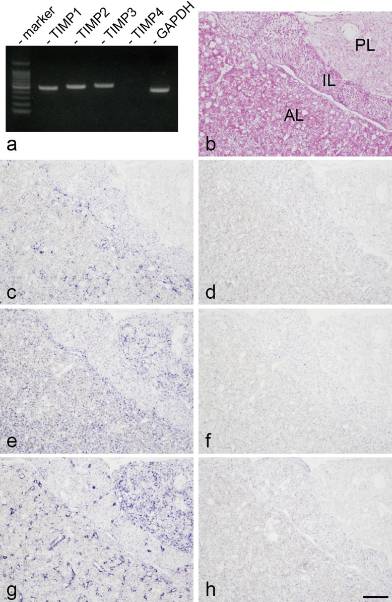
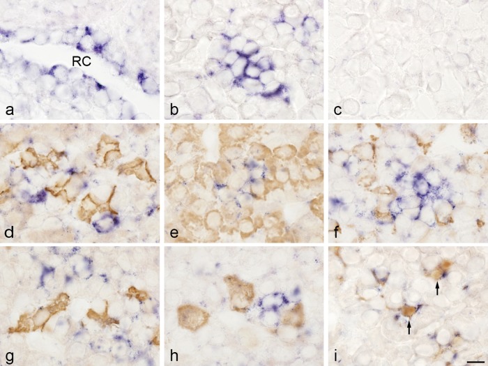
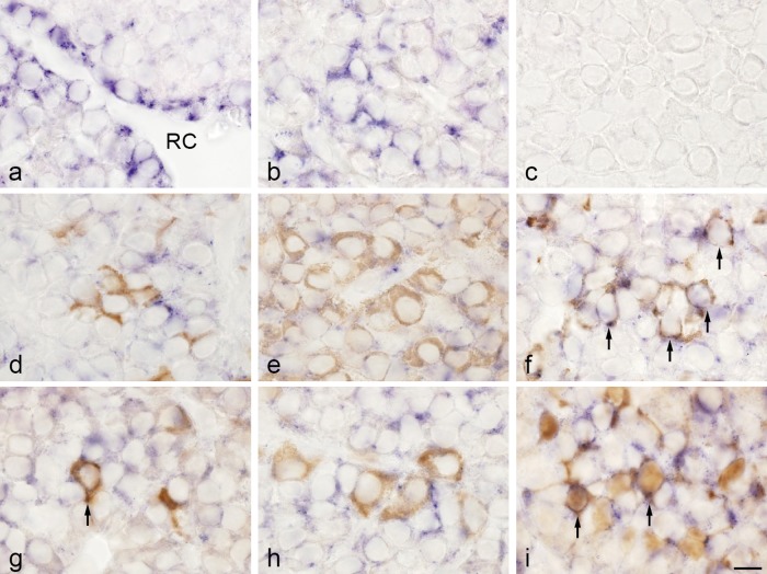
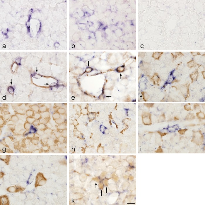
The extracellular matrix (ECM) is important in creating cellular environments in tissues. Recent studies have demonstrated that ECM components are localized in anterior pituitary cells and affect cell activity. Thus, clarifying the mechanism responsible for ECM maintenance would improve understanding of gland function. Tissue inhibitors of metalloproteinases (TIMPs) are endogenous inhibitors of matrix metalloproteinases and participate in ECM degradation. In this study, we investigated whether cells expressing TIMPs are present in rat anterior pituitary gland. Reverse transcription polymerase chain reaction was used to analyze expression of the TIMP family (TIMP1-4), and cells producing TIMPs in the gland were identified by using in situ hybridization. Expression of TIMP1, TIMP2, and TIMP3 mRNAs was detected, and the TIMP-expressing cells were located in the gland. The TIMP-expressing cells were also investigated by means of double-staining with in situ hybridization and immunohistochemical techniques. Double-staining revealed that TIMP1 mRNA was expressed in folliculostellate cells. TIMP2 mRNA was detected in folliculostellate cells, prolactin cells, and thyroid-stimulating hormone cells. TIMP3 mRNA was identified in endothelial cells, pericytes, novel desmin-immunopositive perivascular cells, and folliculostellate cells. These findings indicate that TIMP1-, TIMP2-, and TIMP3-expressing cells are present in rat anterior pituitary gland and that they are involved in maintaining ECM components.

Keywords: anterior pituitary; extracellular matrix; immunohistochemistry; in situ hybridization; tissue inhibitor of metalloproteinases.


Figures

Fig. 1.
Fig. 1.
Expression of tissue inhibitor of metalloproteinase (TIMP) family genes in rat anterior pituitary gland. a: Expression of TIMP family (TIMP1-4) mRNAs in rat anterior pituitary gland, as determined by reverse transcription polymerase chain reaction. Glyceraldehyde-3-phosphate dehydrogenase (GAPDH) mRNA was used as an internal control. b: Hematoxylin and eosin staining of a cryosection of rat anterior pituitary gland (AL: anterior lobe, IL: intermediate lobe, PL: posterior lobe). c, d: In situ hybridization for the TIMP1 antisense probe (c) and sense probe (d). e, f: In situ hybridization for TIMP2 antisense probe (e) and sense probe (f). g, h: In situ hybridization for TIMP3 antisense probe (g) and sense probe (h). Images b–h are consecutive sections. In situ hybridization with NBT/BCIP (blue). Bar=100 μm (b–h).
Fig. 2.
Fig. 2.
Double-staining of TIMP1 mRNA detected by in situ hybridization and hormones and S100 protein detected by immunohistochemistry in rat anterior pituitary gland. a, b: In situ hybridization for TIMP1. TIMP1-expressing cells were observed in the marginal layer (a) surrounding Rathke’s cleft (RC) and in the anterior lobe (b). c: Negative control with sense probe. d–i: In situ hybridization of TIMP1 and immunohistochemistry of adrenocorticotropic hormone (ACTH; d), growth hormone (GH; e), prolactin (f), thyroid-stimulating hormone β-subunit (TSHβ; g), luteinizing hormone β-subunit (LHβ; h), and S100 protein (folliculostellate cells; i). In situ hybridization with NBT/BCIP (blue) and immunostaining with 3,3'-diaminobenzidine (brown). TIMP1 mRNA was colocalized with the S100 protein immunoreaction (arrows). Bar=10 μm (a–i).
Fig. 3.
Fig. 3.
Double-staining of TIMP2 mRNA detected by in situ hybridization and hormones and S100 protein detected by immunohistochemistry in rat anterior pituitary gland. a, b: In situ hybridization for TIMP2. TIMP2-expressing cells were observed in the marginal layer surrounding Rathke’s cleft (RC) (a) and in the anterior lobe (b). c: Negative control with sense probe. d–i: In situ hybridization of TIMP2 and immunohistochemistry of ACTH (d), GH (e), prolactin (f), TSHβ (g), LHβ (h), and S100 protein (folliculostellate cells; i). In situ hybridization with NBT/BCIP (blue) and immunostaining with 3,3'-diaminobenzidine (brown). TIMP2 mRNA was colocalized with the prolactin, TSHβ, and S100 protein immunoreactions (arrows). Bar=10 μm (a–i).
Fig. 4.
Fig. 4.
Double-staining of TIMP3 mRNA detected by in situ hybridization and isolectin B4, desmin, hormones, and S100 protein detected by immunohistochemistry in rat anterior pituitary gland. a, b: In situ hybridization for TIMP3. TIMP3-expressing cells were observed in blood capillaries, perivascular spaces (a), and the anterior lobe (b). c: Negative control with sense probe. d–k: In situ hybridization of TIMP3 and immunohistochemistry of isolectin B4 (endothelial cells; d), desmin (e), ACTH (f), GH (g), prolactin (h), TSHβ (i), LHβ (j), and S100 protein (folliculostellate cells; k). In situ hybridization with NBT/BCIP (blue) and immunostaining with 3,3'-diaminobenzidine (brown). TIMP3 mRNA was colocalized with the isolectin B4, desmin, and S100 protein immunoreactions (arrows). Bar=10 μm (a–k).

Similar articles

Cited by

References

    1. Beaulieu E., Kachra Z., Mousseau N., Delbecchi L., Hardy J. and Béliveau R. (1999) Matrix metalloproteinases and their inhibitors in human pituitary tumors. Neurosurgery 45; 1432–1440; discussion 1440–1441. - PubMed
    1. Brew K., Dinakarpandian D. and Nagase H. (2000) Tissue inhibitors of metalloproteinases: evolution, structure and function. Biochim. Biophys. Acta 1477; 267–283. - PubMed
    1. Chakraborti S., Mandal M., Das S., Mandal A. and Chakraborti T. (2003) Regulation of matrix metalloproteinases: an overview. Mol. Cell. Biochem. 253; 269–285. - PubMed
    1. Fujiwara K., Kikuchi M., Takigami S., Kouki T. and Yashiro T. (2007) Expression of retinaldehyde dehydrogenase 1 in the anterior pituitary glands of adult rats. Cell Tissue Res. 329; 321–327. - PubMed
    1. Fujiwara K., Maekawa F., Kikuchi M., Takigami S., Yada T. and Yashiro T. (2007) Expression of retinaldehyde dehydrogenase (RALDH) 2 and RALDH3 but not RALDH1 in the developing anterior pituitary glands of rats. Cell Tissue Res. 328; 129–135. - PubMed