. 2015;49(6):423-435.
doi: 10.1080/02786826.2015.1040486. Epub 2015 Apr 18.

Profile and Morphology of Fungal Aerosols Characterized by Field Emission Scanning Electron Microscopy (FESEM)

Affiliations

Profile and Morphology of Fungal Aerosols Characterized by Field Emission Scanning Electron Microscopy (FESEM)

Komlavi Anani Afanou et al. Aerosol Sci Technol. 2015.

Abstract

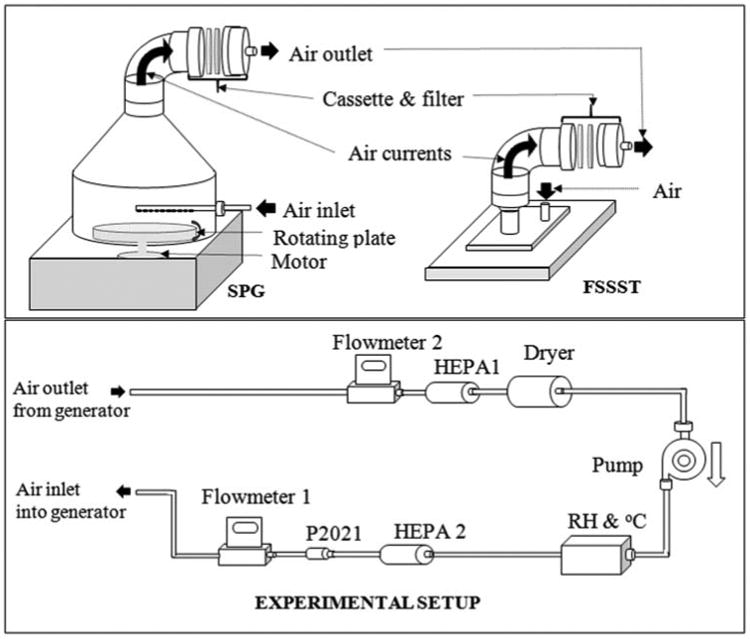
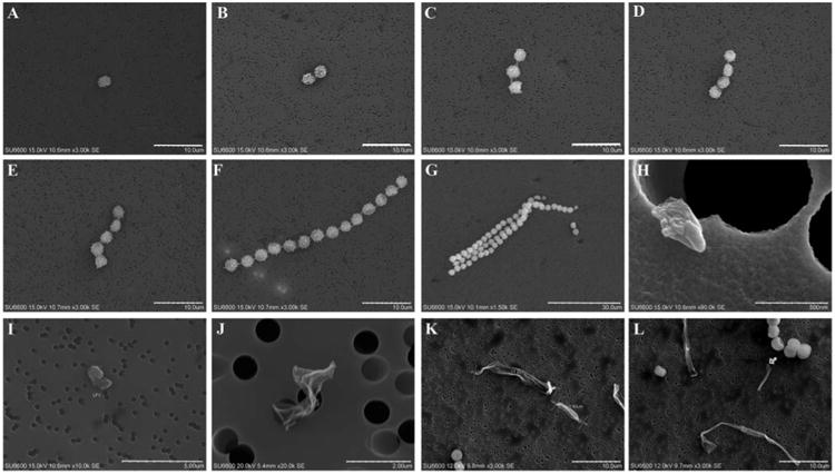
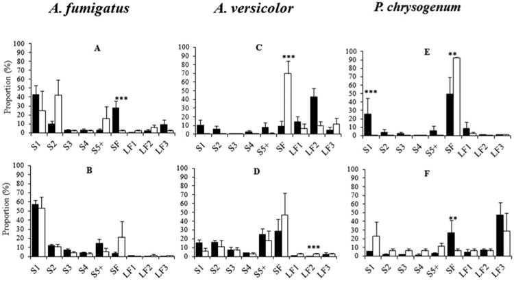
Fungal aerosols consist of spores and fragments with diverse array of morphologies; however, the size, shape, and origin of the constituents require further characterization. In this study, we characterize the profile of aerosols generated from Aspergillus fumigatus, A. versicolor, and Penicillium chrysogenum grown for 8 weeks on gypsum boards. Fungal particles were aerosolized at 12 and 20 L min-1 using the Fungal Spore Source Strength Tester (FSSST) and the Stami particle generator (SPG). Collected particles were analyzed with field emission scanning electron microscopy (FESEM). We observed spore particle fraction consisting of single spores and spore aggregates in four size categories, and a fragment fraction that contained submicronic fragments and three size categories of larger fragments. Single spores dominated the aerosols from A. fumigatus (median: 53%), while the submicronic fragment fraction was the highest in the aerosols collected from A. versicolor (median: 34%) and P. chrysogenum (median: 31%). Morphological characteristics showed near spherical particles that were only single spores, oblong particles that comprise some spore aggregates and fragments (<3.5 μm), and fiber-like particles that regroup chained spore aggregates and fragments (>3.5 μm). Further, the near spherical particles dominated the aerosols from A. fumigatus (median: 53%), while oblong particles were dominant in the aerosols from A. versicolor (68%) and P. chrysogenum (55%). Fiber-like particles represented 21% and 24% of the aerosols from A. versicolor and P. chrysogenum, respectively. This study shows that fungal particles of various size, shape, and origin are aerosolized, and supports the need to include a broader range of particle types in fungal exposure assessment.


Figures

Fig. 1
Fig. 1
Schematic illustration of the aerosolization chambers: Stami particle generator (SPG), fungal spore source strength tester (FSSST) and the experimental set up. One aerosolization chamber is connected to the system set up. Flow at the inlet and outlet of the chamber is measured by flowmeter 1 and 2, respectively. HEPA filter 1 and 2 filter the air to and from the pump, respectively. Constant relative humidity is maintained by the dryer (in line tube with silica beads). P2021 ionizer reduces elctrostatic charges. Reproduced with permission from American Society of Microbiology: Afanou et al. 2014; Applied and Environmental Microbiology, 80(22): 7122–7130.
Fig. 2
Fig. 2
Micrographs of various fungal aerosolized particles. Spore particles (a–f); (a) single spores, (b–g) aggregate of 2, 3, 4, 5, and >5 spores from A. versicolor. Fragment particles (h–l); (h) submicronic fragments: <1 μm; (h–l) large fragments; (i) 1—2 μm fragments; (j) 2–3.5 μm fragments, (k and l) >3.5 μm fragments. Scale bar: 10 μm (a, f, k, and l); 30 μm (g); 0.5 μm (h); 5 μm (i); 2 μm (j).
Fig. 3
Fig. 3
Distribution of mean proportions of particle types aerosolized from A. fumigatus, A. versicolor, and P. chrysogenum cultures; SPG (black columns) and FSSST (white columns) at 12 L min−1 (a, c, and e) and at 20 L min−1 (b, d, and f). S1: Single spores; aggregates of 2 (S2), 3 (S3), 4 (S4), ≥5 (S5) spores. SF: Submicronic fragments, LF1: 1–2 μm fragments, LF2: 2–3.5 μm fragments, and LF3: >3.5 μm fragments. * p ≤ 0.05, ** p ≤ 0.01, *** p ≤ 0.001 (t-test of interaction coefficients between generator and particle types in the centered log-ratio mixed effect regression model of the proportions of particles; all p-values were adjusted for multiple comparisons by the false discovery rate method). Error bars represent standard errors.
Fig. 4
Fig. 4
Distribution of mean proportions of particle types aerosolized from A. fumigatus, A. versicolor, and P. chrysogenum cultures. Airflow 12 L min−1 (black columns) and 20 L min−1 (white columns) with SPG (a, c, and e) and FSSST (b, d, and f). S1: Single spores; aggregates of 2 (S2), 3 (S3), 4 (S4), ≥5 (S5) spores. SF: Submicronic fragments, LF1: 1–2 μm fragments, LF2: 2–3.5 μm fragments and LF3: ≥3.5 μm fragments. * p ≤ 0.05, ** p ≤ 0.01, *** p ≤ 0.001 (t-test of interaction coefficients between airflow and particle types in the centered log-ratio mixed effect regression model of the proportions of particles; all p-values were adjusted for multiple comparisons by the false discovery rate method). Error bars represent standard errors.

References

    1. Adhikari A, Jung J, Reponen T, Lewis JS, DeGrasse EC, Grimsley LF, et al. Aerosolization of Fungi, (1→3)-β-D-Glucans, and Endotoxin from Flood-Affected Materials Collected in New Orleans Homes. Environ Res. 2009;109(3):215–24. doi: 10.1016/j.envres.2008.12.010. - DOI - PMC - PubMed
    1. Afanou KA, Straumfors A, Skogstad A, Nilsen T, Synnes O, Skaar I, et al. Submicronic Fungal Bioaerosols: High-Resolution Microscopic Characterization and Quantification. Appl Environmen Microbiol. 2014;80(22):7122–7130. doi: 10.1128/AEM.01740-14. - DOI - PMC - PubMed
    1. Aitchison J. A concise guide to compositional data analysis. CDA Workshop; Girona: 2003. [Accessed 17 10 2014]. Available at http://www.leg.ufpr.br/lib/exe/fetch.php/pessoais:abtmartins:aconcisegui....
    1. Beckett A, Read ND, Porter R. Variations in Fungal Spore Dimensions in Relation to Preparatory Techniques for Light Microscopy and Scanning Electron Microscopy. J Microsc. 1984;136(1):87–95. doi: 10.1111/j.1365-2818.1984.tb02548.x. - DOI
    1. Benjamini Y. Discovering the False Discovery Rate. J R Stat Soc Series B: Statistical Methodol. 2010;72(4):405–416.