

A Silicon SPECT System for Molecular Imaging of the Mouse Brain
- PMID: 26855557
- PMCID: PMC4743882
- DOI: 10.1109/NSSMIC.2007.4436717
A Silicon SPECT System for Molecular Imaging of the Mouse Brain
Abstract
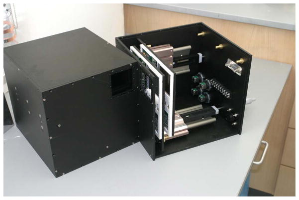
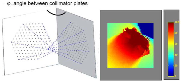
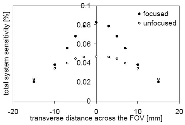
We previously demonstrated the feasibility of using silicon double-sided strip detectors (DSSDs) for SPECT imaging of the activity distribution of iodine-125 using a 300-micrometer thick detector. Based on this experience, we now have developed fully customized silicon DSSDs and associated readout electronics with the intent of developing a multi-pinhole SPECT system. Each DSSD has a 60.4 mm × 60.4 mm active area and is 1 mm thick. The strip pitch is 59 micrometers, and the readout of the 1024 strips on each side gives rise to a detector with over one million pixels. Combining four high-resolution DSSDs into a SPECT system offers an unprecedented space-bandwidth product for the imaging of single-photon emitters. The system consists of two camera heads with two silicon detectors stacked one behind the other in each head. The collimator has a focused pinhole system with cylindrical-shaped pinholes that are laser-drilled in a 250 μm tungsten plate. The unique ability to collect projection data at two magnifications simultaneously allows for multiplexed data at high resolution to be combined with lower magnification data with little or no multiplexing. With the current multi-pinhole collimator design, our SPECT system will be capable of offering high spatial resolution, sensitivity and angular sampling for small field-of-view applications, such as molecular imaging of the mouse brain.
Figures


Similar articles
-
Multi-energy, single-isotope imaging using stacked detectors.Nucl Instrum Methods Phys Res A. 2007 Aug 21;579(1):196-199. doi: 10.1016/j.nima.2007.04.142. Nucl Instrum Methods Phys Res A. 2007. PMID: 19081759 Free PMC article.
-
Multi-pinhole SPECT Imaging with Silicon Strip Detectors.IEEE Trans Nucl Sci. 2009 Jun 16;56(3):646-652. doi: 10.1109/TNS.2009.2012514. IEEE Trans Nucl Sci. 2009. PMID: 20953300 Free PMC article.
-
Collimator design for a multipinhole brain SPECT insert for MRI.Med Phys. 2015 Nov;42(11):667989. doi: 10.1118/1.4934371. Med Phys. 2015. PMID: 26520758
-
The pinhole: gateway to ultra-high-resolution three-dimensional radionuclide imaging.Eur J Nucl Med Mol Imaging. 2007 Feb;34(2):151-61. doi: 10.1007/s00259-006-0248-6. Eur J Nucl Med Mol Imaging. 2007. PMID: 17143647 Review.
-
Improvements in SPECT technology for cerebral imaging.Semin Nucl Med. 1985 Oct;15(4):335-46. doi: 10.1016/s0001-2998(85)80012-x. Semin Nucl Med. 1985. PMID: 3904004 Review.
Cited by
-
Performance Characteristics of Thick Silicon Double-sided Strip Detectors.IEEE Nucl Sci Symp Conf Rec (1997). 2007 Oct-Nov;2007:1656-1660. doi: 10.1109/NSSMIC.2007.4437318. IEEE Nucl Sci Symp Conf Rec (1997). 2007. PMID: 26778911 Free PMC article.
-
Thick Silicon Double-Sided Strip Detectors for Low-Energy Small-Animal SPECT.IEEE Trans Nucl Sci. 2009 Jun 1;56(3):557-564. doi: 10.1109/TNS.2009.2019106. IEEE Trans Nucl Sci. 2009. PMID: 20686626 Free PMC article.
-
A small-animal imaging system capable of multipinhole circular/helical SPECT and parallel-hole SPECT.Nucl Instrum Methods Phys Res A. 2008 Aug 21;594(1):102-110. doi: 10.1016/j.nima.2008.05.061. Nucl Instrum Methods Phys Res A. 2008. PMID: 19701447 Free PMC article.
-
Multi-pinhole collimator design for small-object imaging with SiliSPECT: a high-resolution SPECT.Phys Med Biol. 2009 Jan 21;54(2):207-25. doi: 10.1088/0031-9155/54/2/003. Epub 2008 Dec 16. Phys Med Biol. 2009. PMID: 19088387 Free PMC article.
-
Timing resolution in double-sided silicon photon-counting computed tomography detectors.J Med Imaging (Bellingham). 2023 Mar;10(2):023502. doi: 10.1117/1.JMI.10.2.023502. Epub 2023 Mar 23. J Med Imaging (Bellingham). 2023. PMID: 36969328 Free PMC article.
References
-
- Shokouhi S, Wilson WD, Pham W, Peterson TE. System evaluation for In Vivo imaging of Amyloid beta plaques in a mouse brain using statistical decision theory. IEEE NSS-MIC Conference; 2007.
-
- Wilson DW, Barrett HH, Clarkson EW. Reconstruction of two- and three-dimensional images from synthetic-collimator data. IEEE Trans Med Imaging. 2000;19:412–22. - PubMed
-
- Metzler SD, Bowsher JE, Jaszczak RJ. Geometrical similarities of the Orlov and Tuy sampling criteria and a numerical algorithm for assessing sampling completeness. IEEE Trans Med Imaging. 2003;50:1550–55.
-
- Metzler SD, Bowsher JE, Smith MF, Jaszczak RJ. Analytic determination of pinhole collimator sensitivity with penetration. IEEE Trans Med Imaging. 2001;20:730–41. - PubMed
Grants and funding
LinkOut - more resources
Full Text Sources