. 2016 Jan 19:10:121-35.
doi: 10.2147/OPTH.S90449. eCollection 2016.

Value of optical coherence tomography in the detection of macular pathology before the removal of silicone oil

Affiliations

Value of optical coherence tomography in the detection of macular pathology before the removal of silicone oil

Mohammad Ahmad Rashad et al. Clin Ophthalmol. .

Abstract

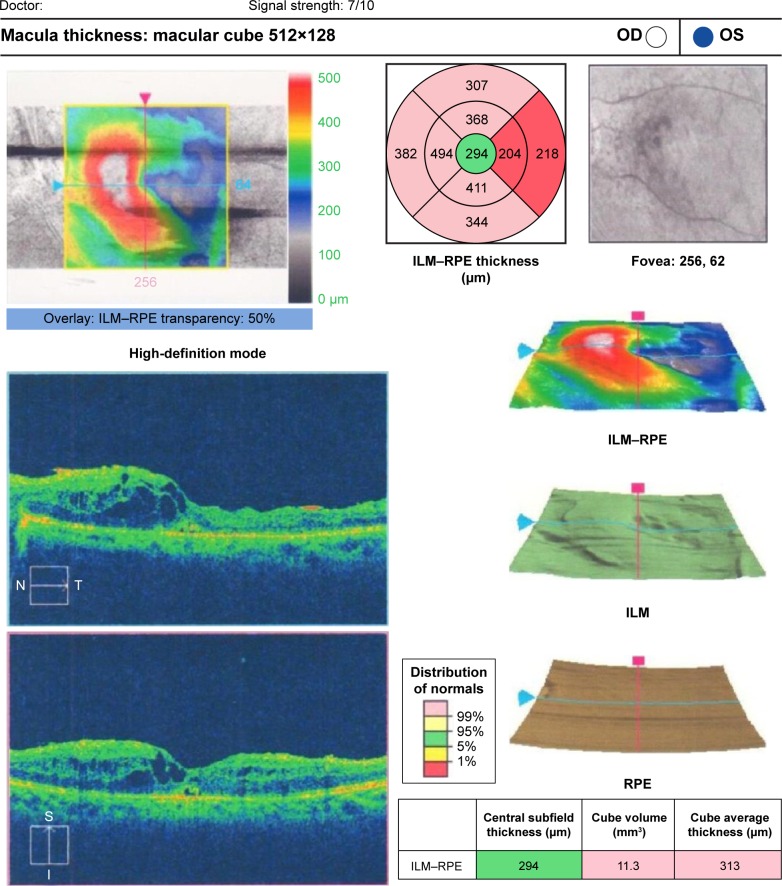
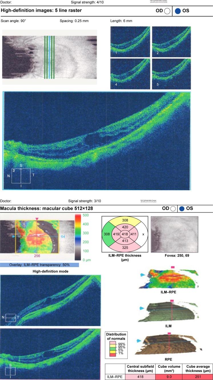
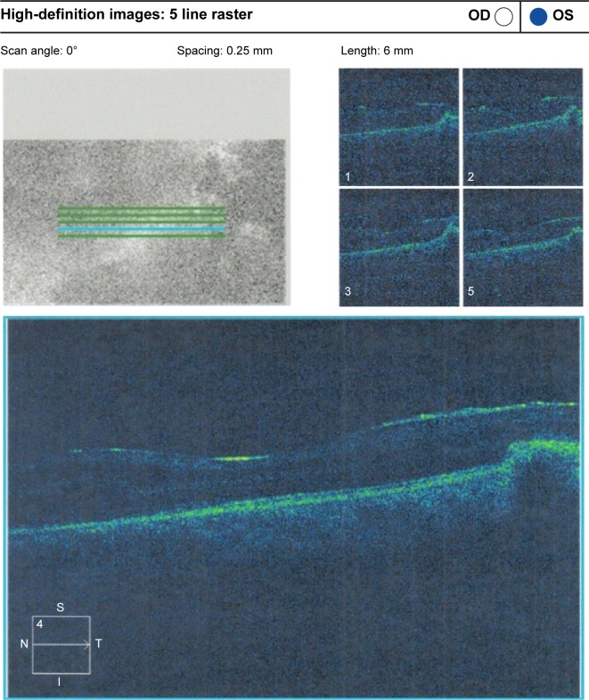
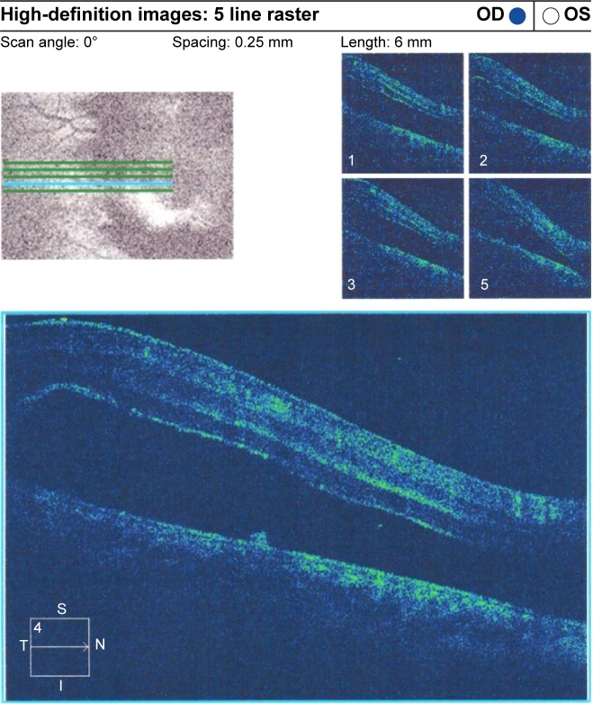
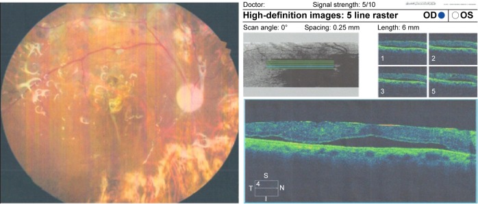
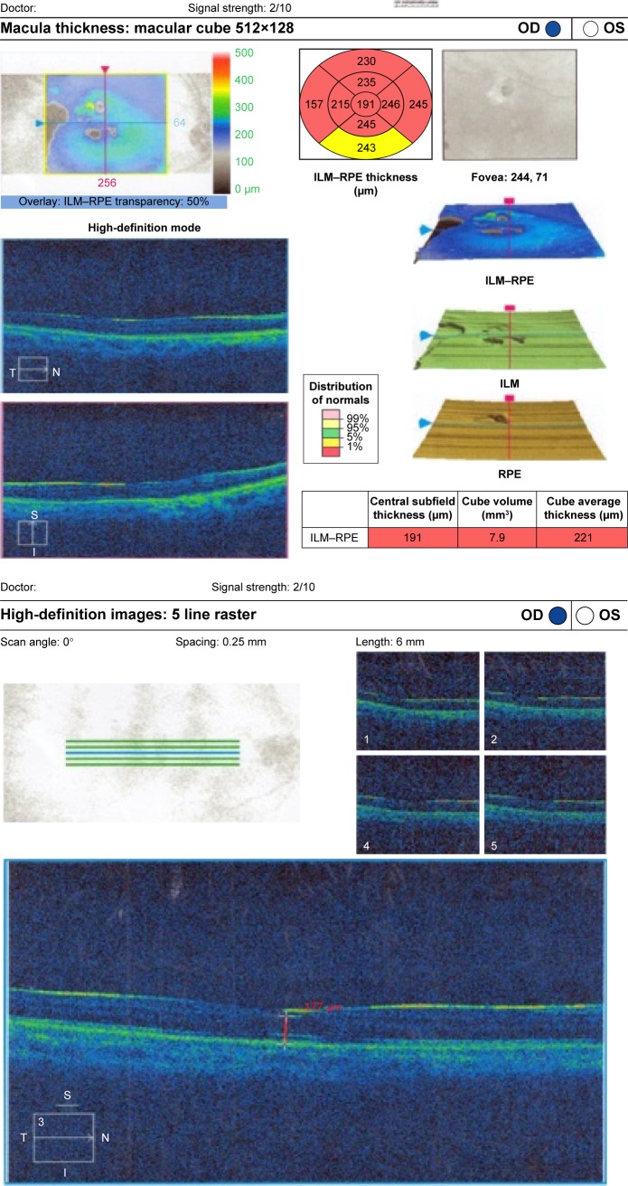
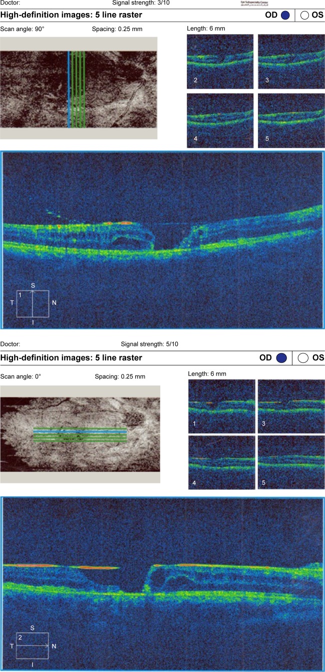
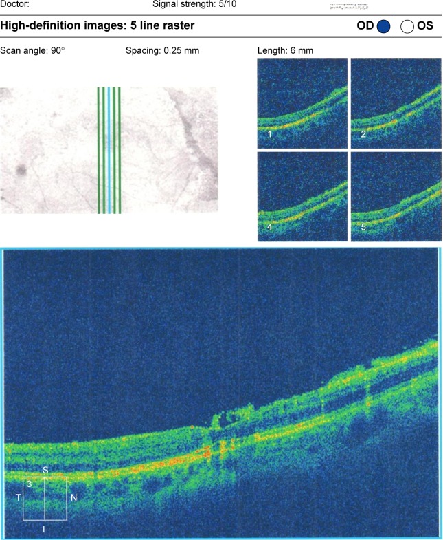
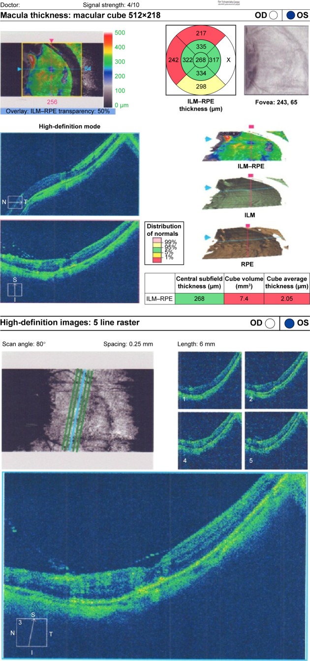
Purpose: To assess the pathological macular changes with optical coherence tomography (OCT) before the removal of silicone oil (SiO) in eyes that had undergone pars plana vitrectomy for complicated forms of retinal detachment (RD).

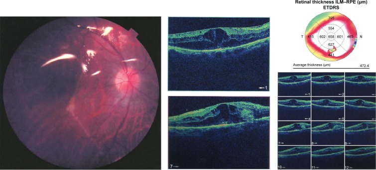
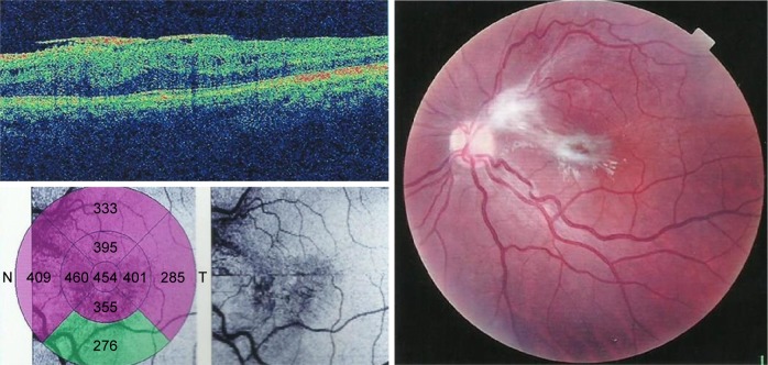
Patients and methods: Subjects included 48 patients (51 eyes) with complicated RD including proliferative vitreoretinopathy, proliferative diabetic retinopathy, recurrent RD, penetrating trauma, uveitis, giant retinal tears, and macular holes. All the eyes had undergone SiO injection. Furthermore, all eyes had been planned for the removal of SiO 6-12 months after the primary surgery. Finally, all eyes had a fundus examination and OCT examination before the silicone oil removal.

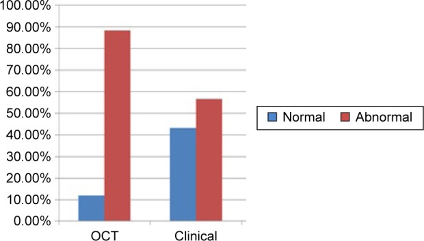
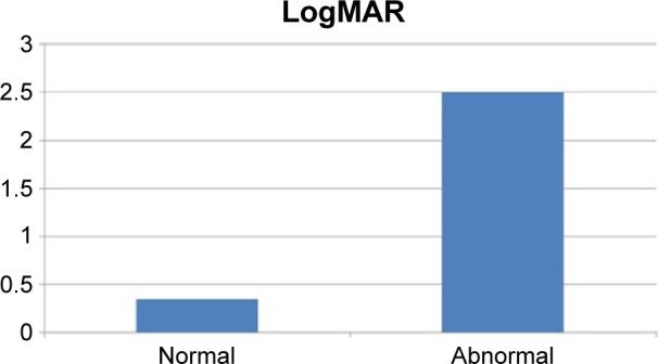
Results: OCT findings indicated epiretinal membrane in 41% of the eyes, macular edema in 17%, macular detachment in 13.5%, macular thinning in 13.5%, macular holes in 10%, and subretinal membranes in 2%. Preoperative OCT was normal in only 12% of the eyes, while a clinical fundus examination was normal in 43% (P<0.001). Eyes with normal OCT had significantly better mean logMAR (0.35) than eyes with pathological changes detected through OCT (1.28; P<0.001). Surgical modifications were made during the removal of SiO in 74.5% of the eyes.

Conclusion: OCT detected significantly more pathological changes than a clinical fundus examination. This had an impact on both surgical step modification during the removal of SiO and predictability of visual outcome after the removal of SiO.

Keywords: optical coherence tomography; pars plana vitrectomy; proliferative diabetic retinopathy; proliferative vitreoretinopathy; silicone oil.


Figures

Figure 1
Figure 1
Diabetic macular edema under SiO. Abbreviations: SiO, silicone oil; OD, right; OS, left; ILM, internal limiting membrane; RPE, retinal pigmented epithelium; t, temporal; S, superior; I, inferior; N, nasal.
Figure 2
Figure 2
(Top) CME under SiO in GRT. (Bottom) CME under SiO in GRT. Abbreviations: CME, cystoid macular edema; SiO, silicone oil; GRT, giant retinal tear; OD, right; OS, left; ILM, internal limiting membrane; RPE, retinal pigmented epithelium; t, temporal; S, superior; I, inferior; N, nasal.
Figure 3
Figure 3
ERM without traction. Abbreviations: ERM, epiretinal membrane; OD, right; OS, left; t, temporal; S, superior; I, inferior; N, nasal.
Figure 4
Figure 4
ERM with traction RD. Abbreviations: ERM, epiretinal membrane; RD, retinal detachment; OD, right; OS, left; t, temporal; S, superior; I, inferior; N, nasal.
Figure 5
Figure 5
Left fundus photo of a diabetic patient with PPV and SiO and OCT of the same patient with shallow macular detachment. Notes: (Left) Colored fundus picture from a diabetic patient who underwent PPV with SiO injection. (Right) OCT raster lines of the same patient showing shallow RD and fine ERM. Abbreviations: PPV, pars plana vitrectomy; SiO, silicone oil; OCT, optical coherence tomography; RD, retinal detachment; ERM, epiretinal membrane; OD, right, OS, left; t, temporal; S, superior; I, inferior; N, nasal.
Figure 6
Figure 6
(Top) Patient fast thickness map. (Bottom) Macular thinning in SiO-filled eye with normal fundus raster lines. Abbreviations: SiO, silicone oil; OD, right; OS, left; t, temporal; S, superior; I, inferior; N, nasal; ILM, internal limiting membrane; RPE, retinal pigmented epithelium.
Figure 7
Figure 7
(Top) Macular hole with macular edema and posterior silicone face. (Bottom) Another raster line of the same patient. Abbreviations: OD, right; OS, left; t, temporal; S, superior; I, inferior; N, nasal.
Figure 8
Figure 8
Raster OC with premacular emulsified SiO in an eye that underwent PPV for recurrent RD. Abbreviations: OC, optical coherence; SiO, silicone oil; PPV, pars plana vitrectomy; RD, retinal detachment; OD, right; OS, left; t, temporal; S, superior; I, inferior; N, nasal.
Figure 9
Figure 9
(Left) Colored fundus photograph of PVRC that underwent PPV. (Right) The same patient with ERM and a subretinal band. Abbreviations: PVR, proliferative retinopathy; PPV, pars plana vitrectomy; ERM, epiretinal membrane; ETDRS, early treatment of diabetic patient study.
Figure 10
Figure 10
OCT detects more abnormalities in the macula than clinical examination. Abbreviation: OCT, optical coherence tomography.
Figure 11
Figure 11
Mean logMAR after the removal of SiO in eyes with normal and abnormal preoperative OCT. Abbreviations: SiO, silicone oil; OCT, optical coherence tomography.
Figure 12
Figure 12
(Left) OCT of a patient with ERM with no tangential traction. (Right) Colored fundus photograph of the same patient with PVR filled with SiO with ERM with tangential traction. Abbreviations: OCT, optical coherence tomography; ERM, epiretinal membrane; PVR, proliferative retinopathy; SiO, silicone oil.
Figure 13
Figure 13
(Top) Fast thickness map improvement of the patient with GRT after pharmacotherapy treatment of CME. (Bottom) Raster line of the same patient. Abbreviations: GRT, giant retinal tear; CME, cystoid macular edema; OD, right; OS, left; t, temporal; S, superior; I, inferior; N, nasal; ILM, internal limiting membrane.

Similar articles

Cited by

References

    1. Huang D, Swanson EA, Lin CP, et al. Optical coherence tomography. Science. 1991;254:1178–1181. - PMC - PubMed
    1. Cibis PA, Becker B, Okun E, Cowan S. The use of liquid silicone in retinal detachment surgery. Arch Ophthalmol. 1962;681:590–599. - PubMed
    1. Scott JD. The treatment of massive vitreous traction by the separation of pre-retinal membranes using liquid silicone oil. Mod Probl Ophthalmol. 1975;15:285–290. - PubMed
    1. Watzke RC. Silicone retinopiesis for retinal detachment: along-term clinical evaluation. Arch Ophthalmol. 1967;77:185–196. - PubMed
    1. Cockerham WD, Schepens CL, Freeman HM. Silicone injection in retinal detachment. Arch Ophthalmol. 1970;83:704–712. - PubMed

LinkOut - more resources