

Langerhans cell histiocytosis: Current concepts in dentistry and case report
- PMID: 26855698
- PMCID: PMC4739360
- DOI: 10.4317/jced.52498
Langerhans cell histiocytosis: Current concepts in dentistry and case report
Abstract
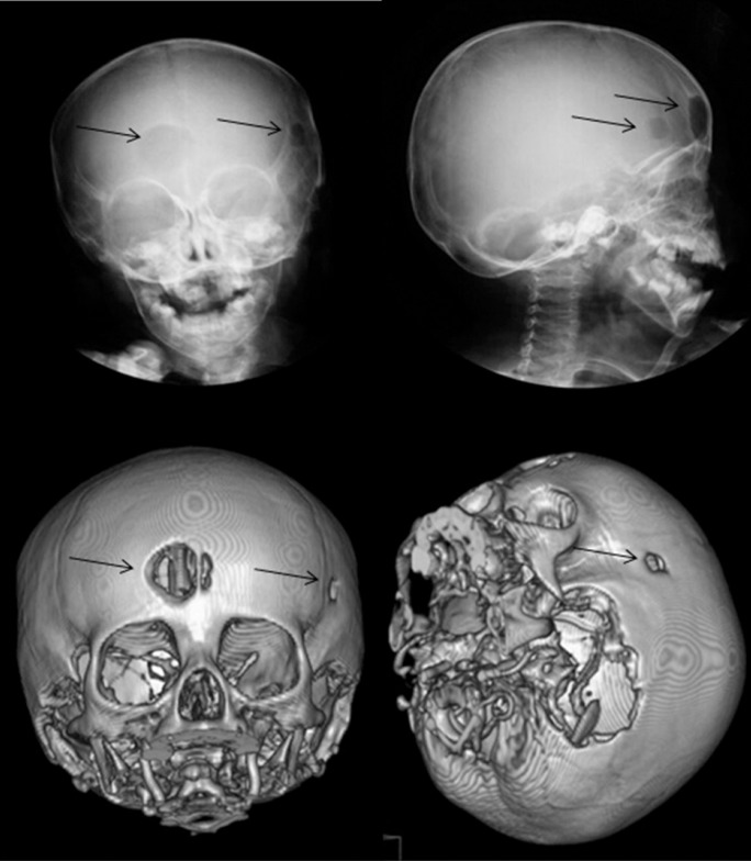
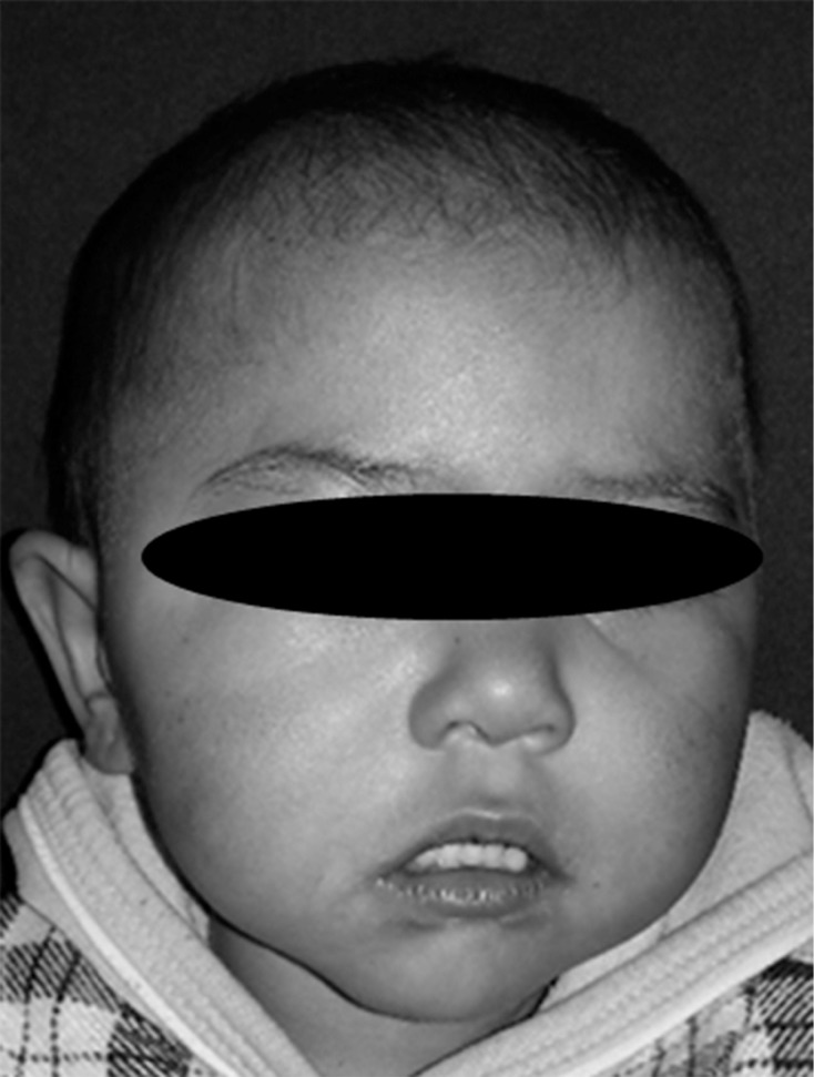
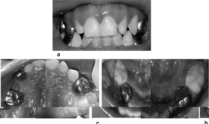
Langerhans cell histiocytosis (LCH), which is a rare granulomatous pediatric disease of unknown etiology, is characterized by the idiopathic proliferation and accumulation of abnormal and clonal Langerhans cells or their marrow precursors, resulting in localized, solitary or multiple destructive lesions. These lesions are most commonly eosinophilic granuloma, which are found in craniofacial bone structures such as the skull and mandible, skin and other organs. In children, the disease has a variable initial presentation, and the clinical course, prognosis and survival are unpredictable. The aims of this report were to present an LCH case in a girl aged 2 years, 8 months and her clinicopathological features, to describe the bucodental management provided, and to discuss special dental considerations of this disease.
Key words: Children, dental management, histiocytosis, Langerhans cells.
Figures
Similar articles
-
Craniofacial Langerhans Cell Histiocytosis: A Rare Case Report.J Coll Physicians Surg Pak. 2021 Oct;31(10):1239-1241. doi: 10.29271/jcpsp.2021.10.1239. J Coll Physicians Surg Pak. 2021. PMID: 34601851
-
Langerhans cell histiocytosis: a diagnostic dilemma.Dent Update. 2012 Dec;39(10):716-8, 720. doi: 10.12968/denu.2012.39.10.716. Dent Update. 2012. PMID: 23367637
-
Co-existence of Langerhans cell histiocytosis and reticulohistiocytosis with initial presentation of skull lesions: A case report.J Cutan Pathol. 2019 Jan;46(1):62-66. doi: 10.1111/cup.13362. Epub 2018 Nov 8. J Cutan Pathol. 2019. PMID: 30251332
-
Langerhans' cell histiocytosis: pathology, imaging and treatment of skeletal involvement.Pediatr Radiol. 2005 Feb;35(2):103-15. doi: 10.1007/s00247-004-1262-0. Epub 2004 Jul 28. Pediatr Radiol. 2005. PMID: 15289942 Review.
-
Multisystem Langerhans' cell histiocytosis (Hand-Schüller-Christian disease) in an adult: a case report and review of the literature.Eur Arch Otorhinolaryngol. 2004 Jul;261(6):326-30. doi: 10.1007/s00405-003-0690-z. Epub 2003 Oct 10. Eur Arch Otorhinolaryngol. 2004. PMID: 14551790 Review.
Cited by
-
Langerhans cell histiocytosis: A diagnostic enigma in the oral cavity.J Oral Maxillofac Pathol. 2021 Mar;25(Suppl 1):S27-S31. doi: 10.4103/jomfp.JOMFP_296_20. Epub 2021 Mar 19. J Oral Maxillofac Pathol. 2021. PMID: 34083966 Free PMC article.
-
Langerhans cell histiocytosis in a pediatric patient: A typical presentation with oral manifestations.Clin Case Rep. 2023 Jul 5;11(7):e7645. doi: 10.1002/ccr3.7645. eCollection 2023 Jul. Clin Case Rep. 2023. PMID: 37426687 Free PMC article.
References
-
- Margo CE, Goldman DR. Langerhans cell histiocytosis. Surv Ophtalmol. 2008;53:332–58. - PubMed
-
- Satter EK, High WA. Langerhans cell histiocytosis: A review of the current recommendations of the Histiocyte Society. Pediatr Dermatol. 2008;25:291–5. - PubMed
-
- Guimarães LF, Dias PF, Janini ME, de Souza IP. Langerhans cell histiocytosis: Impact on the permanent dentition after an 8-year follow-up. J Dent Child. 2008;75:64–8. - PubMed
-
- Badalian-Very G, Vergilio JA, Degar BA, Rodríguez-Galindo C, Rollins BJ. Recent advances in the understanding of Langerhans cell histiocytosis. Brit J Haem. 2011;156:163–72. - PubMed
-
- Madrigal-Martínez-Pereda C, Guerrero-Rodríguez V, Guisado-Moya B, Meniz-García C. Langerhans cell histiocytosis: Literature review and descriptive analysis of oral manifestations. Med Oral Patol Oral Cir Bucal. 2009;14:E222–8. - PubMed
Publication types
LinkOut - more resources
Full Text Sources
Other Literature Sources