

Tension tests on mammalian collagen fibrils
- PMID: 26855757
- PMCID: PMC4686246
- DOI: 10.1098/rsfs.2015.0080
Tension tests on mammalian collagen fibrils
Abstract
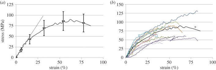
A brief overview of isolated collagen fibril mechanics testing is followed by presentation of the first results testing fibrils isolated from load-bearing mammalian tendons using a microelectromechanical systems platform. The in vitro modulus (326 ± 112 MPa) and fracture stress (71 ± 23 MPa) are shown to be lower than previously measured on fibrils extracted from sea cucumber dermis and tested with the same technique. Scanning electron microscope images show the fibrils can fail with a mechanism that involves circumferential rupture, whereas the core of the fibril stays at least partially intact.
Keywords: collagen fibril; mechanics; nanoscience; nanotechnology; tendon.
Figures
Similar articles
-
Tendon glycosaminoglycan proteoglycan sidechains promote collagen fibril sliding-AFM observations at the nanoscale.J Biomech. 2013 Feb 22;46(4):813-8. doi: 10.1016/j.jbiomech.2012.11.017. Epub 2012 Dec 6. J Biomech. 2013. PMID: 23219277
-
Method to extract minimally damaged collagen fibrils from tendon.J Biol Methods. 2016 Sep 16;3(4):e54. doi: 10.14440/jbm.2016.121. eCollection 2016. J Biol Methods. 2016. PMID: 31453217 Free PMC article.
-
Collagen fibrils in functionally distinct tendons have differing structural responses to tendon rupture and fatigue loading.Acta Biomater. 2016 Sep 15;42:296-307. doi: 10.1016/j.actbio.2016.06.017. Epub 2016 Jun 14. Acta Biomater. 2016. PMID: 27321189
-
STEM/TEM studies of collagen fibril assembly.Micron. 2001 Apr;32(3):273-85. doi: 10.1016/s0968-4328(00)00040-8. Micron. 2001. PMID: 11006507 Review.
-
Collagen structure of tendon relates to function.ScientificWorldJournal. 2007 Mar 30;7:404-20. doi: 10.1100/tsw.2007.92. ScientificWorldJournal. 2007. PMID: 17450305 Free PMC article. Review.
Cited by
-
In vitro fibrillogenesis of tropocollagen type III in collagen type I affects its relative fibrillar topology and mechanics.Sci Rep. 2017 May 3;7(1):1392. doi: 10.1038/s41598-017-01476-y. Sci Rep. 2017. PMID: 28469139 Free PMC article.
-
Strain rate induced toughening of individual collagen fibrils.Appl Phys Lett. 2022 Mar 14;120(11):114101. doi: 10.1063/5.0084054. Epub 2022 Mar 18. Appl Phys Lett. 2022. PMID: 35355883 Free PMC article.
-
In tendons, differing physiological requirements lead to functionally distinct nanostructures.Sci Rep. 2018 Mar 13;8(1):4409. doi: 10.1038/s41598-018-22741-8. Sci Rep. 2018. PMID: 29535366 Free PMC article.
-
A coarse-grained molecular dynamics investigation of the role of mineral arrangement on the mechanical properties of mineralized collagen fibrils.J R Soc Interface. 2023 Jan;20(198):20220803. doi: 10.1098/rsif.2022.0803. Epub 2023 Jan 25. J R Soc Interface. 2023. PMID: 36695019 Free PMC article.
-
Collagen fibrils from both positional and energy-storing tendons exhibit increased amounts of denatured collagen when stretched beyond the yield point.Acta Biomater. 2023 Jan 1;155:461-470. doi: 10.1016/j.actbio.2022.11.018. Epub 2022 Nov 15. Acta Biomater. 2023. PMID: 36400348 Free PMC article.
References
-
- Launey ME, Buehler MJ, Ritchie RO. 2010. On the mechanistic origins of toughness in bone. In Annual review of materials research, vol. 40 (eds Clarke DR, Ruhle M, Zok F), pp. 25–53. Palo Alto, CA: Annual Reviews.
-
- Julkunen P, Wilson W, Isaksson H, Jurvelin JS, Herzog W, Korhonen RK. 2013. A review of the combination of experimental measurements and fibril-reinforced modeling for investigation of articular cartilage and chondrocyte response to loading. Comput. Math. Methods Med. 2013, 326150 (10.1155/2013/326150) - DOI - PMC - PubMed
LinkOut - more resources
Full Text Sources
Other Literature Sources
Miscellaneous
