. 2016 Feb 6:2:2.
doi: 10.1186/s40851-016-0038-3. eCollection 2016.

Amphioxus mouth after dorso-ventral inversion

Affiliations

Amphioxus mouth after dorso-ventral inversion

Takao Kaji et al. Zoological Lett. .

Abstract

Introduction: Deuterostomes (animals with 'secondary mouths') are generally accepted to develop the mouth independently of the blastopore. However, it remains largely unknown whether mouths are homologous among all deuterostome groups. Unlike other bilaterians, in amphioxus the mouth initially opens on the left lateral side. This peculiar morphology has not been fully explained in the evolutionary developmental context. We studied the developmental process of the amphioxus mouth to understand whether amphioxus acquired a new mouth, and if so, how it is related to or differs from mouths in other deuterostomes.

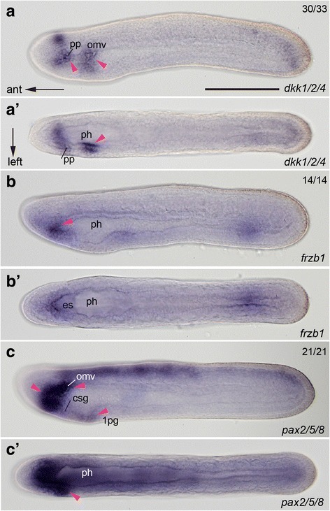
Results: The left first somite in amphioxus produces a coelomic vesicle between the epidermis and pharynx that plays a crucial role in the mouth opening. The vesicle develops in association with the amphioxus-specific Hatschek nephridium, and first opens into the pharynx and then into the exterior as a mouth. This asymmetrical development of the anterior-most somites depends on the Nodal-Pitx signaling unit, and the perturbation of laterality-determining Nodal signaling led to the disappearance of the vesicle, producing a symmetric pair of anterior-most somites that resulted in larvae lacking orobranchial structures. The vesicle expressed bmp2/4, as seen in ambulacrarian coelomic pore-canals, and the mouth did not open when Bmp2/4 signaling was blocked.

Conclusions: We conclude that the amphioxus mouth, which uniquely involves a mesodermal coelomic vesicle, shares its evolutionary origins with the ambulacrarian coelomic pore-canal. Our observations suggest that there are at least three types of mouths in deuterostomes, and that the new acquisition of chordate mouths was likely related to the dorso-ventral inversion that occurred in the last common ancestor of chordates.

Keywords: Coelom; Gill (branchial) slits; Homology of mouth; Hydropore; Lancelet; Nodal-signaling.


Figures

Fig. 1
Fig. 1
Larval asymmetry of amphioxus. a Left lateral view of premetamorphic larva showing streamlined shape. b Magnification of oral region showing mouth and preoral pit on left and gill openings on right side. c Schematic illustration of early knife-shaped larva showing organ formation in anterior region and previously suggested location of mouth opening (arrow). anp; anterior neuropore; ch, notochord; csg, primordial club-shaped gland; es, primordial endostyle; g, gut; lld, left lateral diverticulum (= pp, preoral pit); m, mouth; nc, neurenteric canal; pgs, primary gill slit; rld, right lateral diverticulum (= rc, rostral coelom); 1-10s, 1st-10th somite. Scale bars, 1.0 mm for (a), 0.5 mm for (b)
Fig. 2
Fig. 2
Developmental pattern of oral mesovesicle. ad Mouth formation visualized by pou4 expression. Oral mesovesicle initially connects with first somitocoel (arrowhead in a) and then into pharynx (arrowhead in b). Perforation of mouth occurs through epidermis and remnant of oral mesovesicle (c, d). eg Rendering images of laser confocal microscopic analyses showing expression patterns of pax3/7 and pou4. e13 Anterior to the left. Expression of pax3/7 (orange) in thickened posterior wall of the first somite (arrowheads) from 18 to 30 hpf at 25 °C. f13 Expression of pou4 (orange) in bulged wall of the first somite, which transforms into oral mesovesicle (arrowheads) from 18 to 30 hpf at 25 °C. g13 Outlines of rendering images highlighting expression of two genes. ch, notochord; csg, club-shaped gland; ld, lateral diverticulum; m, mouth; ph, pharynx; pp, preoral pit; 1, 2 s, 1st and 2nd somite; 1, 2sc/L or R, left or right first and second somitocoel; pHn/omv, primordium of Hatschek nephridium/oral mesovesicle complex. Scale bars 50 μm
Fig. 3
Fig. 3
Oral mesovesicle/Hatschek nephridium complex and basal laminae. a, b Fluorescent horizontal sections showing nuclei and immunolabeling for basal laminae. Note dissolution of basal laminae on oral mesovesicle (arrowheads in b). ce Electron micrographs showing transverse sections of developing oral mesovesicle. While primordium of Hatschek nephridium develops basal laminae, oral mesovesicle does not (arrowheads in c). Oral mesovesicle first opens into pharynx without epithelical intercalation (arrowhead in d). Outlet of Hatschek nephridial canal into pharynx (magenta arrowhead in e) and site of oral perforation (green arrowheads in e). f Electron micrographs showing transverse sections of Hatschek nephridium with flagellum and whorl of microvilli from a cyrtopodocyte (cyan arrowhead) and basal laminae (magenta arrowheads). There is no basal lamia around oral mesovesicle (yellow arrowheads). g Magnification of canal of Hatschek nephridium showing two flagella with a whorl of microvilli (cyan arrowheads) and basal lamina between Hatschek nephridium and pharyngeal endoderm (magenta arrowheads). bs, blood sinus; ch, notochord; da, dorsal aorta; ed, epidermis; en, endoderm; Hn, Hatschek nephridium; lld, left lateral diverticulum; omv, oral mesovesicle; ph, pharynx; 2 mm/L, left 2nd myomere;1-2sc/L, left 1st and 2nd somitocoel. Scale bars 20 μm for (a), (b), 5 μm for (cf), and 2 μm for (g)
Fig. 4
Fig. 4
Expression patterns of antagonistic genes to Wnt and pax2/5/8 related to dissolution of basal laminae. ac Left lateral views of knife-shaped larvae. a’–c’ Dorsal views. Most genes are expressed in the region where internal lumens open into the exterior. Arrowheads denote gene expression regions. csg, club-shaped gland; es, endostyle; omv, oral mesovesicle; 1 pg, future first gill; ph, pharynx; pp, preoral pit. Scale bar, 100 μm for all
Fig. 5
Fig. 5
Bilaterally symmetric development of larvae treated with Nodal signaling inhibitor. a Dorsal view showing pitx expression on left side (arrowhead) of untreated 24-hpf larva. b Lack of left-handed pitx expression and symmetrical arrangement of somitocoels (arrowheads) in treated larva. c Mouth opening shown with pou4 expression in control 72-hpf larva. d Disappearance of mouth-related expression of pou4 and complete lack of organs originating from left side and domed pharyngeal floor (arrowhead) in treated larva. e, f Transverse sections showing future oral regions of control larvae at 24 hpf. g, h Transverse sections of treated 24-hpf larva showing bilaterally symmetric internal structures with a pair of rostral coeloms. Levels are comparable between (e) and (g) and (f) and (h), respectively. No oral mesovesicle but somitocoel on left side almost identical to that on right side in treated larvae (compare arrowheads between f and h). i Ventral view of normal pharynx with mouth opening (arrowheads). j, k Ventral views of treated pharynx showing bilaterally symmetric endostyle and club-shaped gland (arrowheads). ik are shown with background of BM purple to clarify morphological details. l, m Immunolabeling for ventral muscles and acetylated tubulin. Left lateral views showing untreated larvae (72 hpf) (l) and SB505124-treated larvae with lack of neurons and oral musculature (66 hpf) (m). anp, anterior neuropore; csg, club-shaped gland; ch, notochord; CNS, central nervous system; es, endostyle; lld, left lateral diverticulum; m, mouth; nt; nerve cord; om, oral muscle; omv, oral mesovesicle; ONR, oral nerve ring; ph, pharynx; pn, peripheral nerve; pp, preoral pit; rc, rostral coelom; 1sc/L or R, first left or right somitocoel. Scale bars 50 μm for (ad), 20 μm for (eh), 100 μm for the others
Fig. 6
Fig. 6
Loss of nephrogenic capability in embryos treated with Nodal signaling inhibitor. aa’ Left lateral (a) and dorsal (a’) views of control hatched-neurula showing lim1/5 expression in posterior wall of left first somite (arrowheads). b, b’ Loss of corresponding expression in treated neurula (circle and arrowheads) viewed left laterally (b) and dorsally (b’). g, gut; lld, left lateral diverticulum; nc, neurenteric canal; 1sc/L or R, first left or right somitocoel. Scale bar, 100 μm for all
Fig. 7
Fig. 7
Expression of bmp2/4 at oral mesovesicle and lack of oral structures in Dorsomophin-treated larvae. a, b Expression of bmp2/4 at future perforation site in oral mesovesicle (arrowhead) at 24 hpf at 25 °C viewed left laterally (a) and dorsally (b). c Control 72-hpf larva having well-developed mouth on the left side. d Muscle fibers of oral musculature immunolabelled with anti-α smooth muscle actin antibody in untreated 72-hpf larva. e, f 72-hpf larva treated with Dorsomorphin reduced size of mouth as a pit (arrowhead in e) and absence of oral musculature (f). g, h Treated larva lacking mouth (g) and oral musculature (h). i A dose-dependent effect of Dorsomorphin on mouth opening treated from 24 to 30 hpf at 25 °C. m, mouth; om, oral musculature; 1, 2 pg, first and second primary gill; pp, preoral pit. Scale bar 100 μm for (a), (b) and the others
Fig. 8
Fig. 8
Schematic drawing of paired gill perforation in amphioxus. a Transverse section showing opening of branchial nephridium into pharynx (arrowhead) at which gill perforation occurs at the dorsal end of atrium (arrow). b Gill opens through coelomic mesothelia on both ectodermal and endodermal sides (arrow). This gill formation is comparable to mouth formation. Redrawn from ref. 50. at, atrium; bn, branchial nephridium; c, coelom; ge, gill epithelium; gp, gill pouch; gs, gill slit; m, myomere; ph, pharynx
Fig. 9
Fig. 9
Mouth formation in deuterostome clade. Ambulacrarians retain ancestral deuterostomic mouth formation, and amphioxus and olfactoreans acquired new methods of mouth formation after dorso-ventral inversion. Amphioxus opens mouth by using coelom and its canal, which has a common origin with ambulacrarian coelomic pore-canals (asterisks). Olfactoreans developed a placode or its equivalent at the anterior extremity to form a stomodaeum

Comment in

Similar articles

Cited by

References

    1. Lee PN, Kumburegama S, Marlow HQ, Martindale MQ, Wikramanayake AH. Asymmetric developmental potential along the animal-vegetal axis in the anthozoan cnidarian, Nematostella vectensis, is mediated by Dishevelled. Dev Biol. 2007;310:169–86. doi: 10.1016/j.ydbio.2007.05.040. - DOI - PubMed
    1. Hejnol A, Martindale MQ. The mouth, the anus, and the blastopore --- open questions about questionable openings. In: Telford MJ, Littlewood DTJ, editors. Animal Evolution: Genomes, Fossils, and Trees. Oxford: Oxford Univ Press; 2009. pp. 33–40.
    1. van den Biggelaar J, Dictus WJAG. Gastrulation in the molluscan embryo. In: Stern CD, editor. Gastrulation: From Cells to Embryo. Cold Spring Harbor: Cold Spring Harbor Laboratory Press; 2004. pp. 63–78.
    1. Hejnol A, Schnabel R. The eutardigrade Thulinia stephaniae has an indeterminate development and the potential to regulate early blastomere ablations. Development. 2004;132:1349–61. doi: 10.1242/dev.01701. - DOI - PubMed
    1. Hejnol A, Martindale MQ. Acoel development indicates the independent evolution of the bilaterian mouth and anus. Nature. 2008;456:382–6. doi: 10.1038/nature07309. - DOI - PubMed

LinkOut - more resources