

Fixation Options for the Volar Lunate Facet Fracture: Thinking Outside the Box
- PMID: 26855830
- PMCID: PMC4742269
- DOI: 10.1055/s-0035-1570739
Fixation Options for the Volar Lunate Facet Fracture: Thinking Outside the Box
Abstract
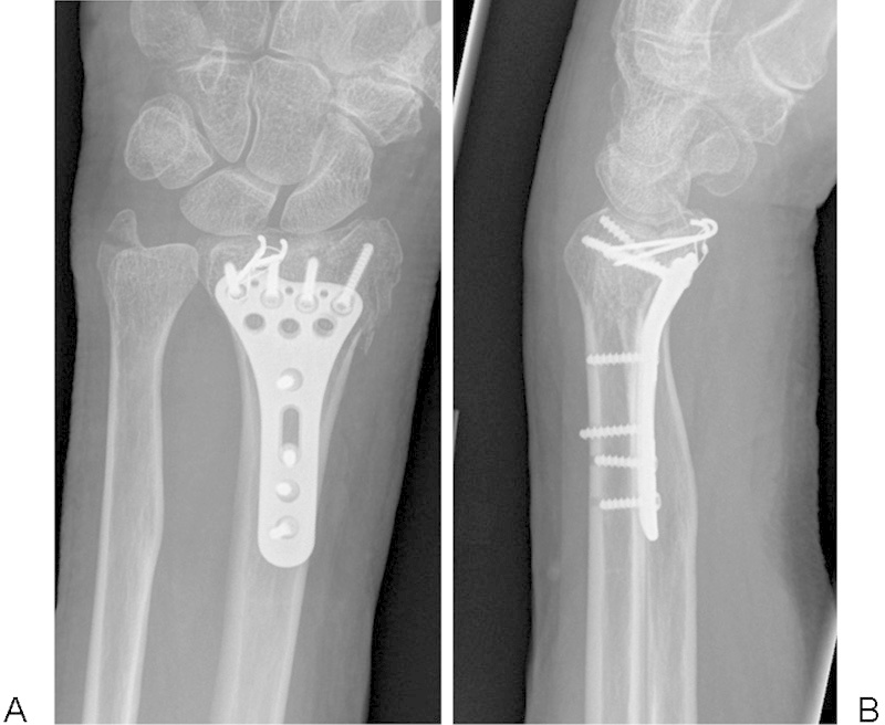
Background Fractures of the distal radius with small volar ulnar marginal fracture fragments are difficult to stabilize with standard volar locking plates. The purpose of this study is to describe alternative techniques available to stabilize these injuries. Materials and Methods Five patients were identified retrospectively with unstable volar lunate facet fracture fragments treated with supplemental fixation techniques. The demographic data, pre- and postoperative radiographic parameters, and early outcomes data were analyzed. The AO classification, preoperative and final postoperative ulnar variance, articular step-off, volar tilt, radial inclination, and teardrop angle were measured. The lunate subsidence and length of the volar cortex available for fixation were measured from the initial injury films. Description of Technique Lunate facet fixation was based on the morphology of the fragment, and stabilization was achieved with headless compression screws in three patients, a tension band wire construct in one, and two cortical screws in another. Results Five patients with a mean age of 58 years (range: 41-82) were included. There were two AO C3.2 and three B3.3 fractures. Preoperative radiographic measurements including radial inclination, tilt, and ulnar variance all improved after surgery and were maintained within normal limits at 3-month follow-up. There was no change in the teardrop angle at final follow-up (70-64 degrees; p = 0.14). None of the patients had loss of fixation or volar carpal subluxation. The mean visual analog scale pain score at 3 months was 1 (range: 0-2). Conclusions The morphology of volar lunate facet fracture fragments is variable, and fixation must be customized to the particular pattern. Small fragments may preclude the use of plates and screws for fixation. These fractures can be managed successfully with tension band wire constructs and headless screws. These low-profile implants may decrease the risk of tendon irritation that might accompany distally placed plates.
Keywords: distal radius; fixation; fracture; lunate facet.
Conflict of interest statement
Figures


References
-
- Harness N G, Jupiter J B, Orbay J L, Raskin K B, Fernandez D L. Loss of fixation of the volar lunate facet fragment in fractures of the distal part of the radius. J Bone Joint Surg Am. 2004;86-A(9):1900–1908. - PubMed
-
- Beck J D, Harness N G, Spencer H T. Volar plate fixation failure for volar shearing distal radius fractures with small lunate facet fragments. J Hand Surg Am. 2014;39(4):670–678. - PubMed
-
- Kitay A, Mudgal C. Volar carpal subluxation following lunate facet fracture. J Hand Surg Am. 2014;39(11):2335–2341. - PubMed
-
- Knirk J L, Jupiter J B. Intra-articular fractures of the distal end of the radius in young adults. J Bone Joint Surg Am. 1986;68(5):647–659. - PubMed
-
- Jupiter J B, Fernandez D L, Toh C L, Fellman T, Ring D. Operative treatment of volar intra-articular fractures of the distal end of the radius. J Bone Joint Surg Am. 1996;78(12):1817–1828. - PubMed
LinkOut - more resources
Full Text Sources
Other Literature Sources
Miscellaneous
