

doi: 10.1055/s-0035-1571186.
Epub 2016 Jan 8.
Fragment-Specific Fixation for Fractures of the Distal Radius
Affiliations
- PMID: 26855832
- PMCID: PMC4742261
- DOI: 10.1055/s-0035-1571186
Item in Clipboard
Fragment-Specific Fixation for Fractures of the Distal Radius
J Wrist Surg.
2016 Mar.
Abstract
This article summarizes the management of distal fractures utilizing Acumed fragment-specific family of plates. No single plate option can address every fracture pattern of the distal radius. These fragment-specific plates are usually adjuncts to allow the surgeon to expand the armamentarium in the management of complex volar and dorsal comminuted distal radius fracture patterns.
Keywords: distal radius; fracture; fragment-specific; ulna; wrist.
Conflict of interest statement
Figures

Acumed (Hillsboro, OR) fragment-specific plates, including the divergent radial styloid plate, volar lunate suture plate, dorsal lunate plate, and dorsal rim buttress plate.
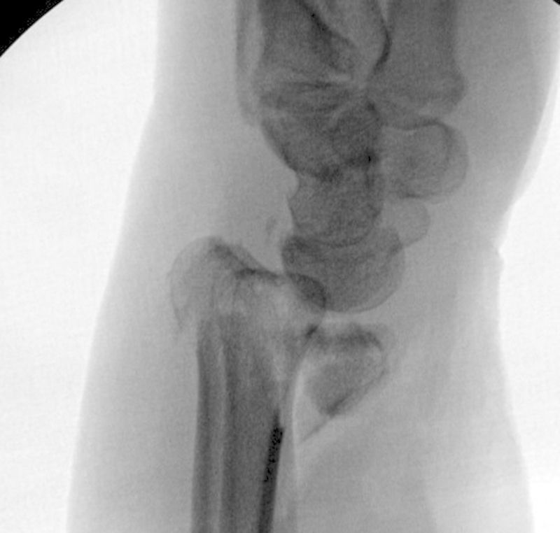
Lateral radiograph of an intraarticular volar Barton's fracture.

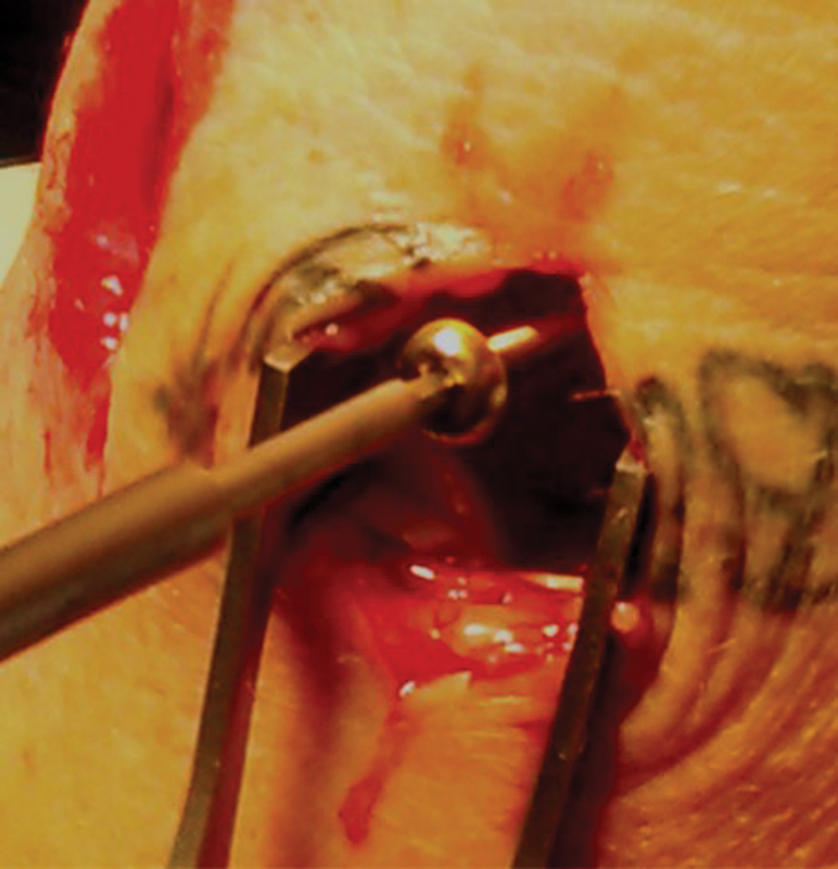
Photograph of the volar locking screw being placed for the Frag-Loc system.
The Frag-Loc being inserted dorsally into the locking screw from the volar plate. Note the incision made to insure the Frag-Loc does not impale any extensor tendons dorsally.
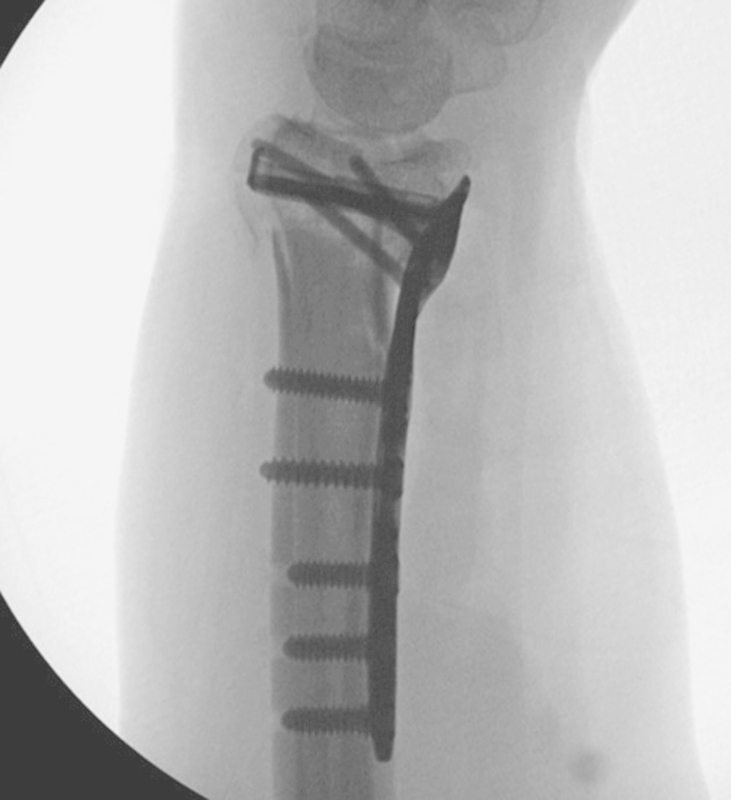
Lateral radiograph showed anatomic joint surface with additional distal support from the Frag-Loc screw.

PA radiograph of a distal intra-articular fracture of the distal radius.

Lateral radiograph showing the distal intra-articular fracture of the distal radius.

Photograph of the Acumed dorsal rim buttress plate with locking guide placed on the dorsal ulnar surface of the radius.

Radiograph showing placement of locking and non-locking screws into the dorsal rim buttress plate.

Posterior/anterior radiograph showing anatomic reduction of the joint surface with stable fixation of both the lunate and radial styloid fragments with a low profile of the dorsal rim plate.

Lateral radiograph showing anatomic reduction of the intra-articular distal radius.

Lateral radiograph showing a dorsal Barton's intra-articular fracture with a radial styloid fracture.

Photograph showing restoration of the dorsal Barton fracture with Acumed's dorsal lunate plate.

Posterior/anterior radiograph showing anatomic reduction with the dorsal lunate plate and an Acutrak screw (Hillsboro, OR) securing the radial styloid fragment. This provided very stable fixation with low profile implants.

Extensively comminuted intra-articular distal radial fracture from a PA radiograph.

Lateral radiograph showing the comminuted intra-articular distal radius fracture.

Sutures being placed through a rotated very distal volar ulnar fragment of the distal radius. The fragment is rotated so the articular surface faces distally.

The sutures are then placed through Acumed's volar lunate suture plate. The sutures are tied after the plate is stabilized to the radial shaft. The plate is used as a buttress to support the small volar fragment.

The patient was extremely large, requiring both the dorsal plate and dorsal lunate plate to support the dorsal comminution.

Posterior/anterior radiographs shows anatomic restoration to the joint surface.

Lateral radiograph demonstrating a plate sandwich with anatomic restoration to the very comminuted intra-articular fracture to the distal radius.

Posterior/anterior radiograph of a radial styloid fracture with metaphyseal comminution.
Stabilization of the radial styloid fracture with the divergent radial styloid plate.

Posterior/anterior radiograph showing anatomic restoration to the joint surface with the divergent radial styloid plate and stabilization of a lunate fracture.

Posterior/anterior radiograph showing a very comminuted fractures involving both the distal radius and ulna.

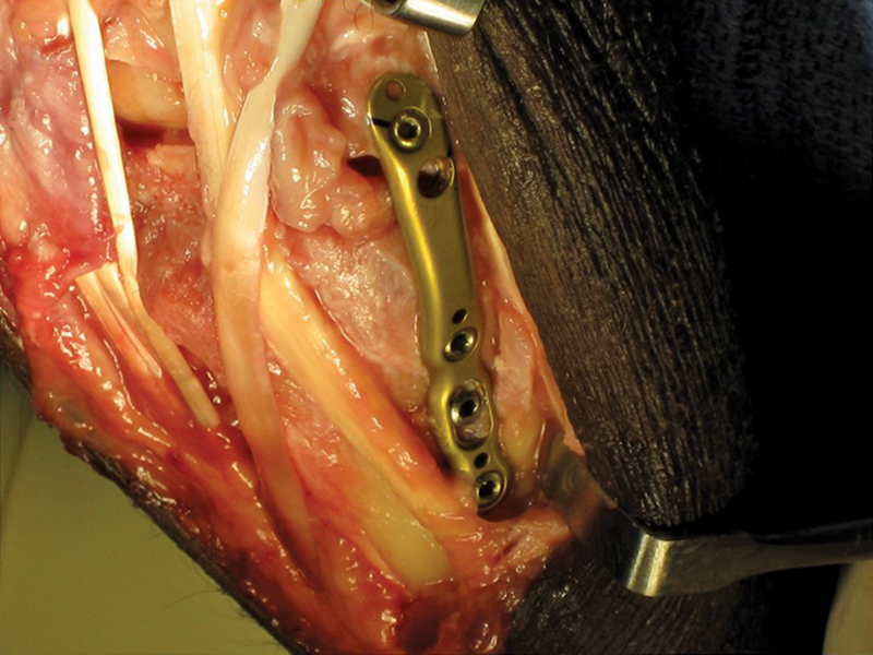
Photograph demonstrating placement of the Acumed volar distal ulnar plate placed on the volar aspect of the distal ulna.
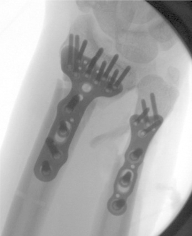
Posterior/anterior radiograph showing anatomic reduction of both of the distal radius and ulna with the volar distal ulna plate.
References
-
- McKay S D, MacDermid J C, Roth J H, Richards R S. Assessment of complications of distal radius fractures and development of a complication checklist. J Hand Surg Am. 2001;26(5):916–922. - PubMed
-
- Catalano L W III, Cole R J, Gelberman R H, Evanoff B A, Gilula L A, Borrelli J Jr. Displaced intra-articular fractures of the distal aspect of the radius. Long-term results in young adults after open reduction and internal fixation. J Bone Joint Surg Am. 1997;79(9):1290–1302. - PubMed
-
- Missakian M L, Cooney W P, Amadio P C, Glidewell H L. Open reduction and internal fixation for distal radius fractures. J Hand Surg Am. 1992;17(4):745–755. - PubMed
-
- Freeland A E, Luber K T. Biomechanics and biology of plate fixation of distal radius fractures. Hand Clin. 2005;21(3):329–339. - PubMed
-
- Orbay J L, Fernandez D L. Volar fixed-angle plate fixation for unstable distal radius fractures in the elderly patient. J Hand Surg Am. 2004;29(1):96–102. - PubMed
LinkOut - more resources
Full Text Sources
Other Literature Sources
