

Application and Efficacy of Super-Magnifying Endoscopy for the Lower Intestinal Tract
- PMID: 26855922
- PMCID: PMC4743732
- DOI: 10.5946/ce.2016.49.1.37
Application and Efficacy of Super-Magnifying Endoscopy for the Lower Intestinal Tract
Abstract
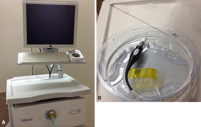
Endoscopy plays a significant role in the diagnosis, management, and surveillance of colorectal cancer (CRC) and inflammatory bowel diseases (IBDs). Moreover, magnifying endoscopy and image-enhanced endoscopy has a crucial role in the clinical setting. Recently, a super-magnifying endoscope has been developed, and two devices, confocal laser endomicroscopy (CLE) and an endocytoscopy system (ECS), which allow in vivo microscopic inspection of the microstructural mucosal features of the gastrointestinal tract, are currently available. Studies on the use of ECS in CRC were reported by a Japanese group. Additionally, a few studies on the use of ECS in IBD have been reported. CLE has been shown to be reliable in assessing the activity of the disease in IBDs in both ulcerative colitis and Crohn's disease. Various published studies evaluated the use of CLE during colonoscopy to distinguish colorectal polyp pathology and neoplasia. However, these studies are heterogeneous, and further evidence is necessary to confirm the efficacy of CLE.
Keywords: Confocal laser endomicroscopy; Endocytoscopy; Magnifying endoscopy; Super-magnifying endoscopy.
Conflict of interest statement
Figures
Similar articles
-
Role of ultra-high definition endoscopy (endomicroscopy and endocytoscopy) and real-time histologic examination in inflammatory bowel disease: Scoping review.Dig Endosc. 2024 Mar;36(3):274-289. doi: 10.1111/den.14659. Epub 2023 Sep 6. Dig Endosc. 2024. PMID: 37573562
-
Confocal laser endomicroscopy for the differential diagnosis of ulcerative colitis and Crohn's disease: a pilot study.Endoscopy. 2015 May;47(5):437-43. doi: 10.1055/s-0034-1391226. Epub 2014 Dec 18. Endoscopy. 2015. PMID: 25521573 Clinical Trial.
-
New endoscopic imaging techniques in surveillance of inflammatory bowel disease.World J Gastrointest Endosc. 2015 Mar 16;7(3):230-6. doi: 10.4253/wjge.v7.i3.230. World J Gastrointest Endosc. 2015. PMID: 25789093 Free PMC article. Review.
-
Confocal laser endomicroscopy in inflammatory bowel diseases: dream or reality?World J Gastroenterol. 2013 Sep 14;19(34):5593-7. doi: 10.3748/wjg.v19.i34.5593. World J Gastroenterol. 2013. PMID: 24039350 Free PMC article.
-
Confocal Laser Endomicroscopy in the Evaluation of Inflammatory Bowel Disease.Inflamm Bowel Dis. 2019 Jul 17;25(8):1302-1312. doi: 10.1093/ibd/izz021. Inflamm Bowel Dis. 2019. PMID: 30877772 Review.
Cited by
-
Microscopic colitis-microbiome, barrier function and associated diseases.Ann Transl Med. 2018 Feb;6(3):39. doi: 10.21037/atm.2017.03.83. Ann Transl Med. 2018. PMID: 29610731 Free PMC article. Review.
References
-
- Oka S, Tanaka S, Chayama K. Detection of nonpolypoid colorectal neoplasia using magnifying endoscopy in colonic inflammatory bowel disease. Gastrointest Endosc Clin N Am. 2014;24:405–417. - PubMed
-
- Santos CE, Pereira-Lima JC, Lopes CV, Malaman D, Parada AA, Salomão AD. Comparative study between MBI (FICE) and magnification chromoendoscopy with indigo carmine in the differential diagnosis of neoplastic and non-neoplastic lesions of the colorectum. Arq Gastroenterol. 2009;46:111–115. - PubMed
-
- Neumann H, Fuchs FS, Vieth M, et al. Review article: in vivo imaging by endocytoscopy. Aliment Pharmacol Ther. 2011;33:1183–1193. - PubMed
-
- Kudo SE, Wakamura K, Ikehara N, Mori Y, Inoue H, Hamatani S. Diagnosis of colorectal lesions with a novel endocytoscopic classification: a pilot study. Endoscopy. 2011;43:869–875. - PubMed
Publication types
LinkOut - more resources
Full Text Sources
Other Literature Sources
