. 2016 Jan 1;2(1):48-62.e3.
doi: 10.1016/j.jcmgh.2015.10.001.

Enterohemorrhagic Escherichia coli reduce mucus and intermicrovillar bridges in human stem cell-derived colonoids

Affiliations

Enterohemorrhagic Escherichia coli reduce mucus and intermicrovillar bridges in human stem cell-derived colonoids

Julie In et al. Cell Mol Gastroenterol Hepatol. .

Abstract

Background and aims: Enterohemorrhagic E. coli (EHEC) causes over 70,000 episodes of foodborne diarrhea annually in the USA. The early sequence of events which precede life-threatening hemorrhagic colitis and hemolytic uremic syndrome are not fully understood due to the initial asymptomatic phase of the disease and the lack of a suitable animal model. The aim of this study was to determine the initial molecular events in the interaction between EHEC and human colonic epithelium.

Methods: Human colonoids derived from adult proximal colonic stem cells were developed into monolayers to study EHEC-epithelial interactions. Monolayer confluency and differentiation were monitored by transepithelial electrical resistance (TER) measurements. The monolayers were apically infected with EHEC and the progression of epithelial damage over time was assessed using biochemical and imaging approaches.

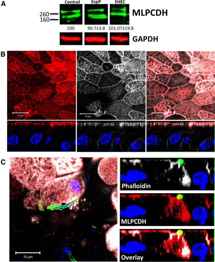
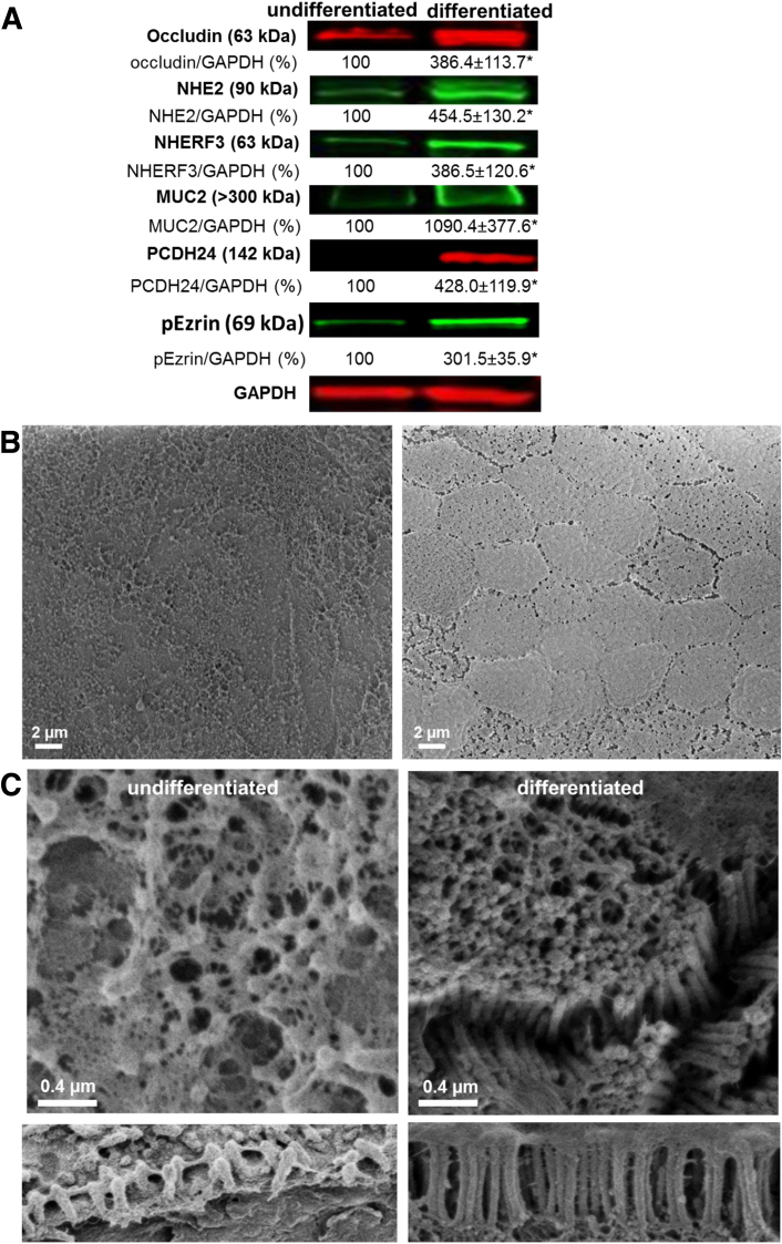
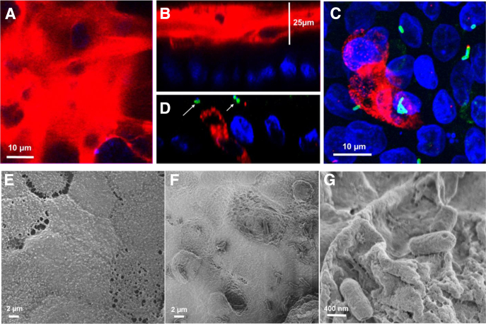
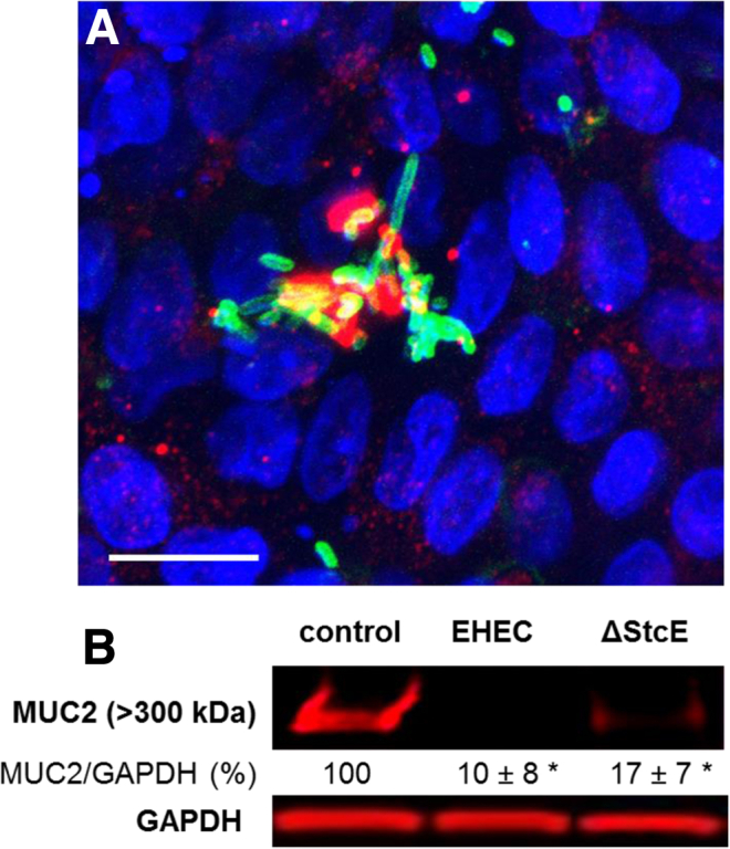
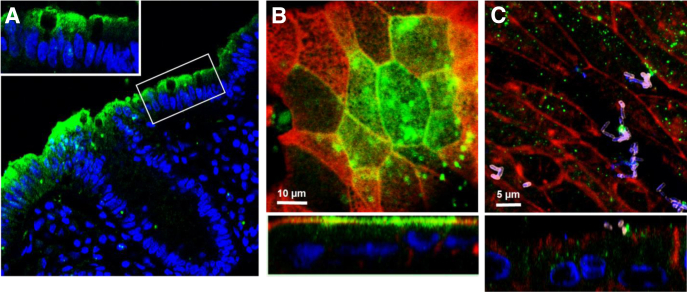
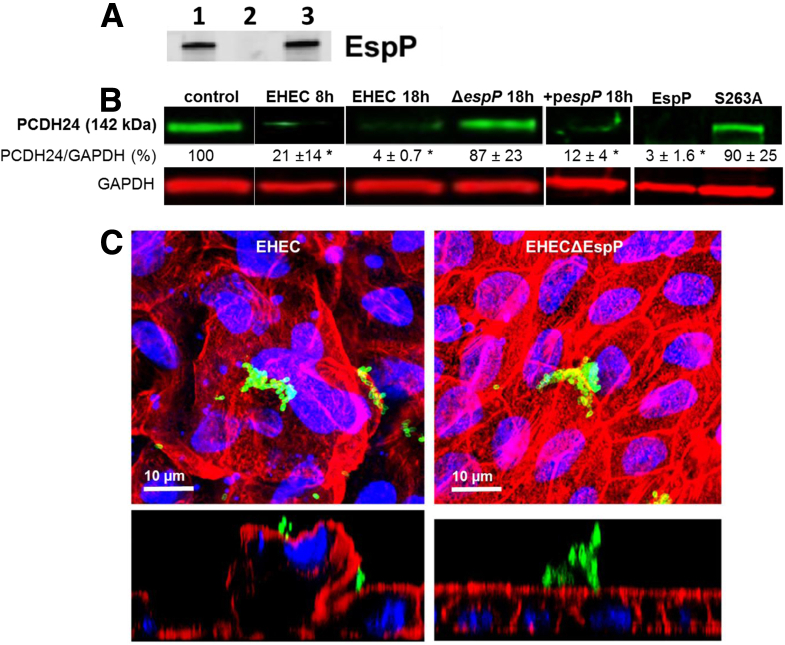
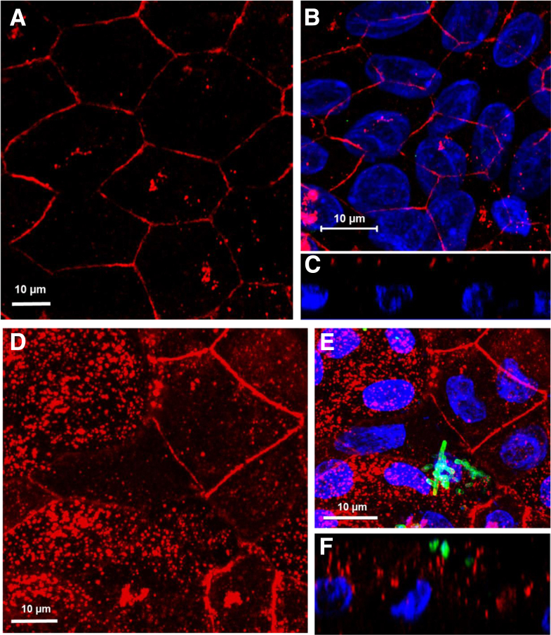
Results: Human colonoid cultures recapitulate the differential protein expression patterns characteristic of the crypt and surface colonocytes. Mucus-producing differentiated colonoid monolayers are preferentially colonized by EHEC. Upon colonization, EHEC forms characteristic attaching and effacing lesions on the apical surface of colonoid monolayers. Mucin 2, a main component of colonic mucus, and protocadherin 24 (PCDH24), a microvillar resident protein, are targeted by EHEC at early stages of infection. The EHEC secreted serine protease, EspP, initiates brush border damage through PCDH24 reduction.

Conclusions: Human colonoid monolayers are a relevant pathophysiological model which allows the study of early molecular events during enteric infections. Colonoid monolayers provide access to both apical and basolateral surfaces, thus providing an advantage over 3D cultures to study host-pathogen interactions in a controllable and tractable manner. EHEC reduces colonic mucus and affects the brush border cytoskeleton in the absence of commensal bacteria.

Keywords: human colonoid monolayers; intestinal organoids; microvillar effacement; serine protease EspP.


Figures

Supplementary Figure 1
Differentiation results in a decrease of the stem cell marker leucine-rich repeat containing G protein-coupled receptor 5 (LGR5). Relative LGR5 expression was measured by semiquantitative polymerase chain reaction in three different human colonoid lines (undifferentiated and differentiated for 5 days). LGR5 was normalized to RN18S and relative expression levels from the three colonoid lines were averaged. Average LGR5 expression was 877.4 ± 157.9 and 9.9 ± 3.5 in undifferentiated and differentiated, respectively. *Statistically significant decrease of LGR5 expression, P < .05, n = 5.
Supplementary Figure 2
Confirmation of protocadherin 24 (PCDH24) reduction by EspP. PCDH24 expression is significantly decreased with enterohemorrhagic Escherichia coli (EHEC) or EHECΔstcE, but minimally with EHECΔespP infection. Lower molecular weight, or cleaved, forms of PCDH24 are not detected, possibly due to the lack of antigen recognition by the antibody used.
Supplementary Figure 3
Mucin-like protocadherin (MLPCDH), a protocadherin 24 (PCDH24) binding partner, is mislocalized, but protein expression level does not change upon enterohemorrhagic Escherichia coli (EHEC) infection. Representative immunoblot of MLPCDH in control and infected human colonoid monolayers. The MLPCDH expression level does not change upon EHEC infection (up to 18 hours) or treatment with recombinant EspP (10 μM, 60 minutes). (A) Representative image of a differentiated colonoid monolayer immunostained for MLPCDH (red, left panel), actin (phalloidin, white, middle panel), and overlayed in the right panel; DNA (Hoechst, blue). Note that MLPCDH is mainly on the apical surface and overlaps with the actin pattern. (B) Representative image of an EHEC-infected differentiated colonoid monolayer immunostained for actin (phalloidin, white), MLPCDH (red), EHEC antigen (green), and DNA (Hoechst, blue). Overlay in the XY plane is shown on the left panel. XZ planes (right panels) show that MLPCDH becomes mislocalized from the BB into the cytosol.
Figure 1
Figure 1
Removal of Wnt3A induces differentiation of colonoids. (A) The relative amount of each marker in both undifferentiated and differentiated colonoids normalized to GAPDH as shown in Western blots. *Statistically significant increase, P < .05, n = 3. Protein expression changes show transition to surface colonocyte population upon differentiation. (B) Scanning electron microscope (SEM) images of undifferentiated and differentiated colonoid monolayers at low magnification show the uniform mucus-covered brush border (BB) in differentiated monolayers compared with the sparse and uneven BB of the undifferentiated monolayers. (C) Scanning electron microscope images of undifferentiated and differentiated colonoid monolayers at higher magnification show distinct changes in the BB and microvillar height (lower panel) induced by differentiation.
Figure 2
Figure 2
Enterohemorrhagic Escherichia coli (EHEC) preferentially colonize differentiated colonoid monolayers. (A) Representative confocal three-dimensional projections from images of undifferentiated and (B) differentiated human colonoid monolayers (HCM) infected with 106 EHEC for 8 hours. Phalloidin (red), DNA (blue), EHEC antigen (green). Bars: 10 μM. (C) Representative confocal three-dimensional projection from images of differentiated jejunal monolayers infected with 106 EHEC for 8 hours. Very few EHEC were found attached or associated. Phalloidin (red), EHEC antigen (green). Bar: 10 μM. (D) Quantification of EHEC or EHECΔespA bacteria associated with undifferentiated and differentiated colonic monolayers. Twenty separate fields (each measuring 50 × 50 μm) were counted for EHEC and averaged. EHECΔespA were counted in 12 separate fields (each measuring 50 × 50 μm) and averaged (n = three independent experiments). **P < .001; *P < .05.
Figure 3
Figure 3
Enterohemorrhagic Escherichia coli (EHEC) infection targets the mucus layer in human colonoids. (A, B) Differentiated human colonoid monolayers (HCM) express a thick (>25 μm) inner mucus layer primarily composed of MUC2 (red). DNA (blue). (A) XY projection; (B) XZ projection. (C, D) EHEC infection for 4 hours reduces thickness of the extracellular mucin 2 (MUC2)–positive mucus layer. Remaining MUC2 (red) is intracellular in goblet cells. Arrows point to attached EHEC (green). (C) XY projection; (D) XZ projection . (E–G) Scanning electron microscope images of HCM (E) uninfected and (F, G) EHEC-infected after 6 hours. EHEC infection leads to microvillar effacement and apical membrane perturbation (F) and EHEC attachment to the remaining glycocalyx (G).
Figure 4
Figure 4
Enterohemorrhagic Escherichia coli (EHEC) ΔstcE is able to degrade the human colonoid monolayers (HCM) mucus layer. (A) Representative confocal three-dimensional projection from images of EHECΔstcE-infected HCM shows that EHECΔstcE infection does not prevent the extracellular mucin 2 (MUC2)–positive mucus layer reduction. Remaining MUC2 (red) resides intracellularly in goblet cells. EHEC antigen (green), DNA (blue). Bar: 10 μM. (B) MUC2 expression normalized to GAPDH is significantly decreased (*P < .05) in EHEC and EHECΔstcE infected colonoids compared with uninfected.
Figure 5
Figure 5
Enterohemorrhagic Escherichia coli (EHEC) forms attaching and effacing (A/E) lesions and actin pedestals while also remodeling the basolateral membranes. (A) Scanning electron microscope and (B) transmission electron microscope (TEM) images of EHEC-infected differentiated human colonoid monolayers (HCM) after 8 hours. EHEC effaces the microvilli and attaches via actin pedestals (arrows, inset of A). (C) EHEC attaches to the apical surface of HCM via F-actin pedestals. Note the overlapping actin with EHEC (arrow). F-actin (red), EHEC antigen (green), DNA (blue). (D, E) TEM images of (D) uninfected differentiated HCM and (E) EHEC-infected differentiated HCM show that apical infection with EHEC causes membrane perturbation and actin remodeling of both apical and basolateral membranes. Note that the apical tight junctions were less affected by EHEC infection.
Figure 6
Figure 6
EspP significantly decreases the amount of protocadherin 24 (PCDH24) and remodels the brush border (BB). (A) Representative image of normal human colonic tissue immunostained for PCDH24 shows that PCDH24 is localized to the apical surface of colonocytes and to the brush border. Inset shows a zoomed-in section of the BB. PCDH24 (green), DNA (blue). (B, C) PCDH24 expression on (B) uninfected and (C) enterohemorrhagic Escherichia coli (EHEC)–infected (8 hours) human colonoid monolayers (HCM). PCDH24 is localized to the apical membrane of uninfected HCM and in intracellular puncta of infected HCM. PCDH24 (green), F-actin (red), EHEC antigen (pink), DNA (blue). XY projection (top panels), XZ projection (bottom panels).
Figure 7
Figure 7
EspP significantly decreases the amount of protocadherin 24 (PCDH24) and remodels the BB. (A) EspP production was determined by Western blot analysis using a polyclonal EspP antiserum. EspP was detected in overnight cultures of wild-type enterohemorrhagic Escherichia coli (EHEC) (lane 1), and the EHECΔespP strain complemented with a plasmid expressing espP (lane 3), but not in the culture of the EHECΔespP strain (lane 2). (B) Expression of PCDH24 normalized to GAPDH is quantitated after infection by wild-type EHEC, mutant strain EHECΔespP, or complemented espP mutant strain (pespP), and after treatment (5 hours) by purified recombinant EspP or EspP S263A. *Statistically significant decrease compared with control (P < .05, n ≥ 3 per each condition). (C) Infection with wild-type EHEC, but not EHECΔespP, causes apical membrane perturbation and actin remodeling of the BB. F-actin (red), EHEC antigen (green), DNA (blue).
Figure 8
Figure 8
Enterohemorrhagic Escherichia coli (EHEC) causes redistribution of occludin from tight junctions to cytosolic puncta. (AC) Occludin is localized to the lateral membrane of uninfected differentiated human colonoid monolayers (HCM). Occludin (red), DNA (blue). (A, B) XY projection; (C) XZ projection. (DF) EHEC infection (18 hours) of HCM causes occludin redistribution to cytosolic puncta and significant loss from the tight junction. Occludin (red), EHEC antigen (green), DNA (blue). (D, E) XY projection; (F) XZ projection.

Similar articles

Cited by

References

    1. Karmali M.A., Gannon V., Sargeant J.M. Verocytotoxin-producing Escherichia coli (VTEC) Vet Microbiol. 2010;140:360–370. - PubMed
    1. Nguyen Y., Sperandio V. Enterohemorrhagic E. coli (EHEC) pathogenesis. Front Cell Infect Microbiol. 2012;2:90. - PMC - PubMed
    1. Goldwater P.N., Bettelheim K.A. Treatment of enterohemorrhagic Escherichia coli (EHEC) infection and hemolytic uremic syndrome (HUS) BMC Med. 2012;10:12. - PMC - PubMed
    1. Pacheco A.R., Sperandio V. Shiga toxin in enterohemorrhagic E. coli: regulation and novel anti-virulence strategies. Front Cell Infect Microbiol. 2012;2:81. - PMC - PubMed
    1. Kovbasnjuk O., Mourtazina R., Baibakov B. The glycosphingolipid globotriaosylceramide in the metastatic transformation of colon cancer. Proc Natl Acad Sci USA. 2005;102:19087–19092. - PMC - PubMed