. 2016 Feb 9:6:21499.
doi: 10.1038/srep21499.

Changing expression profiles of lncRNAs, mRNAs, circRNAs and miRNAs during osteoclastogenesis

Affiliations

Changing expression profiles of lncRNAs, mRNAs, circRNAs and miRNAs during osteoclastogenesis

Ce Dou et al. Sci Rep. .

Abstract

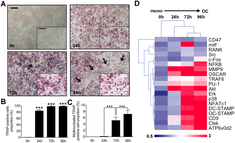
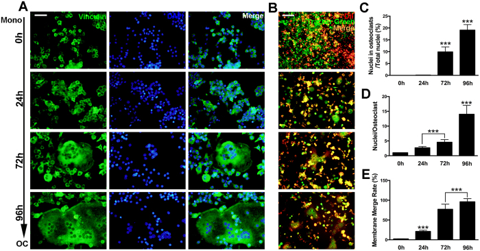
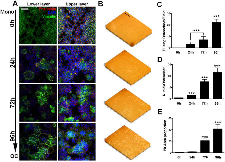
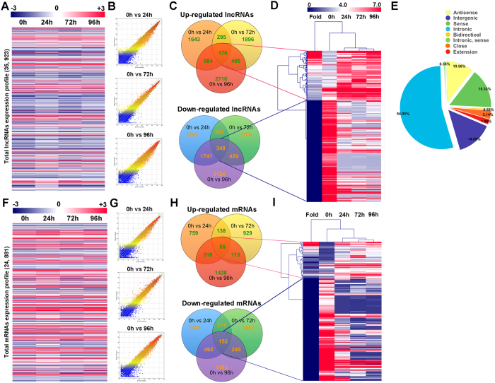
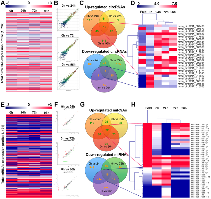
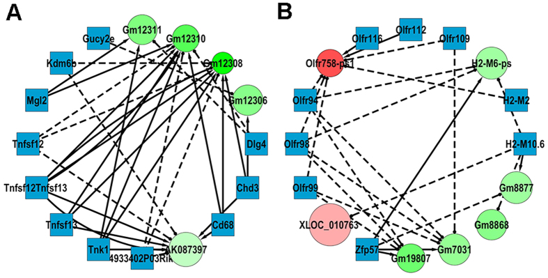
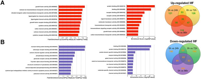
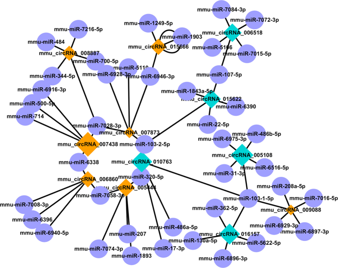
Bone is a dynamic organ continuously undergoing shaping, repairing and remodeling. The homeostasis of bone is maintained by the balance between osteoblastic bone formation and osteoclastic bone resorption. Osteoclasts (OCs) are specialized multinucleated cells derived from hematopoietic stem cells (HSCs) or monocytes/macrophage progenitor cells. There are different stages during osteoclastogenesis, and one of the most important steps to form functional osteoclasts is realized by cell-cell fusion. In our study, microarray was performed to detect the expression profiles of lncRNA, mRNA, circRNA and miRNA at different stages during osteoclastogenesis of RAW264.7 cells. Often changed RNAs were selected and clustered among the four groups with Venn analysis. The results revealed that expressions of 518 lncRNAs, 207 mRNAs, 24 circRNAs and 37 miRNAs were often altered at each stage during OC differentiation. Gene ontology (GO) and Kyoto Encyclopedia of Genes and Genomes (KEGG) biological pathway analysis were performed to predict the functions of differentially expressed lncRNAs and co-expressed potential targeting genes. Co-expression networks of lncRNA-mRNA and circRNA-miRNA were constructed based on the correlation analysis between the differentially expressed RNAs. The present study provided a systematic perspective on the potential function of non-coding RNAs (ncRNAs) during osteoclastogenesis.


Figures

Figure 1
Figure 1. Osteoclasts differentiation and specific genes expression change at different stages.
(A) Representative images of TRAP stain of osteoclasts cultured from RAW264.7 cells induced with RANKL (100 ng/mL) and M-CSF (50 ng/mL) for 0 h, 24 h, 72 h and 96 h. The upper scale bar represents 200 μm and the lower scale bar represents 50 μm. Multinucleated cells were highlighted by black arrows. (B) Quantitative analysis of TRAP positive cell proportion of at each stage during the differentiation. (C) Proportion of TRAP positive cells with more than three nuclei (three included) in total TRAP positive cells at each differentiation stage. (D) Osteoclasts specific and fusogenic genes expression level alterations from 0 h (monocytes) to 96 h (mature osteoclasts).The data in the figures represent the averages ± SD. *p < 0.05, **p < 0.01, and ***p < 0.001 based on one-way ANOVA.
Figure 2
Figure 2. Osteoclasts formation and fusion at different stages.
(A) Representative images of FAK stain of osteoclasts cultured from RAW264.7 cells induced with RANKL (100 ng/mL) and M-CSF (50 ng/mL) for 0 h, 24 h, 72 h and 96 h. The scale bar represents 100 μm. (B) Representative images of fusion assay. Cells were induced with RANKL (100 ng/mL) and M-CSF (50 ng/mL) for 0 h, 24 h, 72 h and 96 h. The scale bar represents 100 μm. (C) Quantification of the total number of nuclei in osteoclasts over the total number of nuclei. (D) Average nuclei number of osteoclasts of FAK stain at different stages. (E) Membrane merge rate of osteoclasts from fusion assay at each differentiation stage. The data in the figures represent the averages ± SD. *p < 0.05, **p < 0.01, and ***p < 0.001 based on one-way ANOVA.
Figure 3
Figure 3. Osteoclasts sealing zone formation and bone resorption activity at different stages.
(A) Representative images of FAK stain of osteoclasts at different stages during differentiation. Sealing zone was observed at lower layers with confocal microscopy. The scale bar represents 20 μm. (B) Representative images of pit formation assay. Cells were induced with RANKL (100 ng/mL) and M-CSF (50 ng/mL)for 0 h, 24 h, 72 h and 96 h.The scale bar represents 800 μm. (C) Number of fusing osteoclasts at different stages. (D) Average nuclei number of osteoclasts at different stages in FAK stain. (E) Quantification of pit area formation in (B). The data in the figures represent the averages ± SD. *p < 0.05, **p < 0.01, and ***p < 0.001 based on one-way ANOVA.
Figure 4
Figure 4. Expression profiles of lncRNAs and mRNAs during osteoclasts differentiation and fusion.
(A) The cluster heat map of all lncRNAs expression at different stages during osteoclastogenesis from microarray data. (B) Scatter plots showing differentially expressed lncRNAs between osteoclasts at different stages and monocytes (0 h). (C) Often differentially expressed lncRNAs between osteoclasts at different stages and monocytes (0 h). (D) Hierarchical clustering showing often up and down regulated lncRNAs among the four groups. (E) lncRNAs categories and distribution in the microarray. (F) The cluster heat map of all mRNAs expression at different stages during osteoclastogenesis from microarray data. (G) Scatter plots showing differentially expressed mRNAs between osteoclasts at different stages and monocytes (0 h). (H) Often differentially expressed mRNAs between osteoclasts at different stages and monocytes (0 h). (I) Hierarchical clustering showing often up and down regulated mRNAs among the four groups.
Figure 5
Figure 5. Expression profiles of circRNAs and miRNAs during osteoclasts differentiation and fusion.
(A) The cluster heat map of all circRNAs expression at different stages during osteoclastogenesis from microarray data. (B) Scatter plots showing differentially expressed circRNAs between osteoclasts at different stages and monocytes (0 h). (C) Often differentially expressed circRNAs between osteoclasts at different stages and monocytes (0 h). (D) Hierarchical clustering showing often up and down regulated circRNAs among the four groups. (E) The cluster heat map of all miRNAs expression at different stages during osteoclastogenesis from microarray data. (F) Scatter plots showing differentially expressed miRNAs between osteoclasts at different stages and monocytes (0 h). (G) Often differentially expressed miRNAs between osteoclasts at different stages and monocytes (0 h). (H) Hierarchical clustering showing often up and down regulated miRNAs among the four groups.
Figure 6
Figure 6. Construction of the lncRNA-mRNA co-expression network.
Circle nodes represent lncRNA and square nodes represent mRNAs. Red color and green color represent up and down regulation respectively. The shade darkness of red and green represents fold change of lncRNAs. The size of circle represents p-value with larger size owing smaller p-value. Solid lines represent positive relationship and dash lines represent negative relationship. (A,B) Detailed presentation of two sub-networks in the dashed box in Fig. S6.
Figure 7
Figure 7. GO analysis of the biological function of lncRNA co-expression genes.
(A) Often up regulated GO MF terms for the difference lncRNAs co-expression genes were analyzed. Top 10 often up regulated GO terms ranked by fold enrichment and enrichment score were shown. (B) Often down regulated GO MF terms for the difference lncRNAs co-expression genes were analyzed. Top 10 often down regulated GO terms ranked by fold enrichment and enrichment score were shown.
Figure 8
Figure 8. Construction of the circRNA-miRNA co-expression network.
Construction of the circRNA-miRNA co-expression network. Diamond nodes represent circRNAs and purple circle nodes represent miRNAs. Yellow color and blue color represents up and down regulation respectively. The size of diamonds represents fold change of circRNAs with larger size owing higher fold change.

References

    1. Boyle W. J., Simonet W. S. & Lacey D. L. Osteoclast differentiation and activation. Nature 423, 337–342 (2003). - PubMed
    1. Boyce B. F., Rosenberg E., de Papp A. E. & Duong le T. The osteoclast, bone remodelling and treatment of metabolic bone disease. European journal of clinical investigation 42, 1332–1341 (2012). - PubMed
    1. Yasuda H. et al. Osteoclast differentiation factor is a ligand for osteoprotegerin/osteoclastogenesis-inhibitory factor and is identical to TRANCE/RANKL. Proceedings of the National Academy of Sciences of the United States of America 95, 3597–3602 (1998). - PMC - PubMed
    1. Bozec A. et al. T cell costimulation molecules CD80/86 inhibit osteoclast differentiation by inducing the IDO/tryptophan pathway. Science translational medicine 6, 235ra260 (2014). - PubMed
    1. Kim N. et al. Osteoclast differentiation independent of the TRANCE-RANK-TRAF6 axis. The Journal of experimental medicine 202, 589–595 (2005). - PMC - PubMed

Publication types