. 2016 Apr;22(4):623-35.
doi: 10.1261/rna.054411.115. Epub 2016 Feb 8.

Cooling-induced SUMOylation of EXOSC10 down-regulates ribosome biogenesis

Affiliations

Cooling-induced SUMOylation of EXOSC10 down-regulates ribosome biogenesis

John R P Knight et al. RNA. 2016 Apr.

Abstract

The RNA exosome is essential for 3' processing of functional RNA species and degradation of aberrant RNAs in eukaryotic cells. Recent reports have defined the substrates of the exosome catalytic domains and solved the multimeric structure of the exosome complex. However, regulation of exosome activity remains poorly characterized, especially in response to physiological stress. Following the observation that cooling of mammalian cells results in a reduction in 40S:60S ribosomal subunit ratio, we uncover regulation of the nuclear exosome as a result of reduced temperature. Using human cells and an in vivo model system allowing whole-body cooling, we observe reduced EXOSC10 (hRrp6, Pm/Scl-100) expression in the cold. In parallel, both models of cooling increase global SUMOylation, leading to the identification of specific conjugation of SUMO1 to EXOSC10, a process that is increased by cooling. Furthermore, we define the major SUMOylation sites in EXOSC10 by mutagenesis and show that overexpression of SUMO1 alone is sufficient to suppress EXOSC10 abundance. Reducing EXOSC10 expression by RNAi in human cells correlates with the 3' preribosomal RNA processing defects seen in the cold as well as reducing the 40S:60S ratio, a previously uncharacterized consequence of EXOSC10 suppression. Together, this work illustrates that EXOSC10 can be modified by SUMOylation and identifies a physiological stress where this regulation is prevalent both in vitro and in vivo.

Keywords: 40S subunits; RNA exosome; SUMOylation; cold shock; rRNA processing.


Figures

FIGURE 1.
FIGURE 1.
40S subunit abundance is reduced by mild hypothermia due to defects in rRNA processing. (A) HEK293 cells were maintained at 37°C (red line) or 32°C for 24 h (blue line), then ribosomal subunits separated by sucrose density ultracentrifugation. The relative change in 40S:60S ratio is annotated beside the trace, which is the average of three independent experiments ±SEM. P = 0.043. (B) Cells treated as in A were analyzed using lysis buffer and density gradients containing 25 μM EDTA to dissociate polysomes. Relative change in 40S:60S ratio is annotated from three independent replicates ±SEM. P = 0.024. (C) Schematic representation of pre-rRNAs in human cells from the initial 47S/45S transcript to the mature 18S, 5.8S, and 28S rRNAs. The internal (ITS) and external (ETS) transcribed spacers are indicated. (D) Total RNA extracted from HEK293 cells incubated for up to 24 h at 32°C was size separated and used in Northern blotting using [32P] labeled oligonucleotide probes complementary to specific sequences of rRNAs. 18S and 28S were detected by Northern blot (NB). (E) Quantification of changes in rRNAs during mild hypothermia of HEK293 cells shown in D. The 41S values are taken using the ITS1 probe. The average rRNA abundance from three biological repeats is shown ±SEM. (*) P < 0.05, (**) P < 0.01. (F) Total RNA was extracted at the indicated times into the chase of a pulse-chase rRNA labeling with [32P] orthophosphate in cooled and control HEK293 cells. Size-separated RNA visualized by methylene blue (MB) staining and autoradiography. (G) Quantification of rRNA abundances in F plotted relative to the 180 min 37°C sample for each rRNA, which has been set to one. n = 3. 41S (P = 0.037), 21S (P = 0.034), 12S (P = 0.040), 7S (P = 0.002).
FIGURE 2.
FIGURE 2.
Global SUMOylation is increased by mild hypothermia. HEK293 cells were incubated at 32°C for 4 or 24 h or maintained at 37°C, then whole cell lysates were analyzed by Western blotting. Quantification of the abundance of >150 kDa high molecular weight (HMW) SUMO1, SUMO2/3 and ubiquitin protein conjugates from three independent experiments are given below each blot. Values are the averages ± SEM. Both SUMO1 and SUMO2/3 HMW conjugates are significantly increased—P = 0.028 and 0.041, respectively.
FIGURE 3.
FIGURE 3.
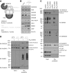
EXOSC10 is SUMOylated and its expression reduced by cooling. (A) Schematic representation of the mammalian exosome. EXOSC1–3 constitutes the cap, part of the catalytically inert core when combined with EXOSC4–9. EXOSC10 binds the core at the cap and contains exonucleolytic activity—denoted by a black circle. Dis3 binds to the base of the core and contains two RNase site (black circles). The subcellular distribution of each component is given. (B) HEK293 cells were incubated at 32°C for either 4 or 24 h and compared to cells maintained at 37°C. Lysates from these cells were analyzed by Western blot for the expression of the proteins shown. eEF2 T56-P, RBM3, and CIRP induction are indicative of the reduction in temperature. β-actin is used as a loading control. (C) His6-tagged constructs encoding SUMO1 or SUMO2 were transiently transfected into HEK293 cells, which were then cultured at 37°C for 48 h. Cells were then lysed and His6-tagged proteins precipitated and analyzed by Western blotting as the His6 pulldown, compared to whole cell lysates termed the input. Conjugated proteins are annotated. (D) His6-SUMO1 was expressed at 37°C or 32°C as in C and conjugated proteins precipitated. Western blotting for EXOSC10 abundance in the pulldowns, using RanGAP1 as a loading control for a precipitated protein. Conjugated proteins are annotated. Dashed lines indicate removal of lanes.
FIGURE 4.
FIGURE 4.
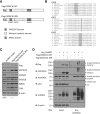
SUMOylation of EXOSC10 reduces its expression. (A) Schematic representation of Flag-tagged wild-type and a mutant construct encoding EXOSC10. Lysine (K) residues mutated to arginine (R) are shown. The gray boxed area shows the N-terminal Flag tag with endogenous domains of EXOSC10 also annotated. (B) Sequence conservation of putative SUMO sites was analyzed using ClustalW with the SUMOylated lysine highlighted—including SUMOylation motif where present. (C) The abundance of Flag-tagged EXOSC10 was analyzed by Western blot 48 h following transfection into HEK293 cells subsequently incubated at 32°C for 24 h. The expression of endogenous EXOSC10 at 37°C is included to illustrate the loss of protein upon cooling. (*) Indicates a nonspecific band. (D) His6-tagged SUMO1 was expressed alone or with each Flag-tagged EXOSC10 construct for 48 h, followed by precipitation of His6-tagged proteins. The control lane containing no construct was transfected with empty pcDNA. Annotations show the conjugated proteins in the pulldowns.
FIGURE 5.
FIGURE 5.
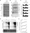
EXOSC10 knockdown recapitulates the cooling-induced defects in ribosome biogenesis. (A) HEK293 cells were transfected with siRNAs as shown then cultured for a further 48 h. Cooled cells were transferred to 32°C for the final 24 h. Lysates were prepared for SDS-PAGE and the expression of the indicated proteins determined by Western blot. The induction of eEF2 T56-P, RBM3, and CIRP is used to confirm the cooling response and β-tubulin is used as a loading control. (B) Cells were treated in parallel to A, then total RNA size separated and analyzed by Northern blotting. (C) Quantification of the abundance of pre-rRNAs shown in B following cooling or siRNA suppression of EXOSC10. The abundance of each pre-rRNA is compared to mock treatment at 37°C, which is set to one (gray line). The data show the average of three biological repeats ±S.E.M. (†) Indicates a P-value approaching significance (P = 0.061 for both). (D) HEK293 cells were transfected with EXOSC10 siRNA 1, then after 48 h at 37°C cells labeled with [32P] orthophosphate and the incorporation into nascent RNA analyzed by agarose formaldehyde electrophoresis. Methylene blue (MB) staining is used as a loading control, with calculated abundances normalized to this. The pre-rRNA and rRNA forms attributed to each band are annotated. (E) Quantification of the abundance of the indicated pre-rRNAs standardized to the abundance of 47/45S pre-rRNA at 0 min for each pre-rRNA.
FIGURE 6.
FIGURE 6.
EXOSC10 maintains the 40S:60S ratio and rate of global protein synthesis. (A) Cells were transfected with EXOSC10 siRNAs and maintained at 37°C for 48 h before analysis by sucrose density ultracentrifugation to quantify free subunit and polysome abundance. (B) The free 40S:60S ratio relative to mock transfected cells at 37°C was calculated from A. Values shown are the average of two independent experiments ±SEM. (C) Cells were treated as in A, the incorporation of [35S] label into nascent protein measured by scintillation counting and expressed relative to mock transfection at 37°C. The values are the average of three biological replicates ±SEM.
FIGURE 7.
FIGURE 7.
In vivo cooling induces ribosome biogenesis defects and an altered 40S:60S ratio. (A) Western blotting from mouse lung tissue from control or 5′AMP cooled mice were analyzed for the abundance of the indicated proteins. The change in SUMO isoforms and EXOSC10 are indicated, standardized to β-actin. Values are the average ±SEM where n = 3. SUMO1 HMW conjugates are significantly increased, P = 0.021. EXOSC10 is significantly reduced P = 0.026. (B) Total RNA from the hippocampi of cooled or control treated mice was extracted and analyzed by Northern blot. Methylene blue (MB) staining is used to visualize the abundance of mature rRNAs. (C) Pre-rRNAs quantified and expressed relative to total RNA, defined as the cumulative abundance of 18S and 28S rRNAs. Values for the three replicates shown are averaged and expressed ±SEM. 47S/45S (P = 0.034), 29S (P = 0.049), 21S (P = 0.040). (D) Hippocampal lysates generated from cooled or control mice were analyzed by sucrose density ultracentrifugation in the absence of EDTA. Average values for the areas calculated for the 40S and 60S are shown and converted into the 40S:60S ratio. Data are the average of three mice ±SEM. P = 0.049.

Similar articles

Cited by

References

    1. Al-Fageeh MB, Marchant RJ, Carden MJ, Smales CM. 2006. The cold-shock response in cultured mammalian cells: harnessing the response for the improvement of recombinant protein production. Biotechnol Bioeng 93: 829–835. - PubMed
    1. Anderson JS, Parker RP. 1998. The 3′ to 5′ degradation of yeast mRNAs is a general mechanism for mRNA turnover that requires the SKI2 DEVH box protein and 3′ to 5′ exonucleases of the exosome complex. EMBO J 17: 1497–1506. - PMC - PubMed
    1. Becker J, Barysch SV, Karaca S, Dittner C, Hsiao HH, Berriel Diaz M, Herzig S, Urlaub H, Melchior F. 2013. Detecting endogenous SUMO targets in mammalian cells and tissues. Nat Struct Mol Biol 20: 525–531. - PubMed
    1. Cole SE, LaRiviere FJ, Merrikh CN, Moore MJ. 2009. A convergence of rRNA and mRNA quality control pathways revealed by mechanistic analysis of nonfunctional rRNA decay. Mol Cell 34: 440–450. - PMC - PubMed
    1. Danno S, Nishiyama H, Higashitsuji H, Yokoi H, Xue JH, Itoh K, Matsuda T, Fujita J. 1997. Increased transcript level of RBM3, a member of the glycine-rich RNA-binding protein family, in human cells in response to cold stress. Biochem Biophys Res Commun 236: 804–807. - PubMed

Publication types