. 2016 Mar;18(3):465-80.
doi: 10.1016/j.jcyt.2015.12.007.

Ex vivo virotherapy with myxoma virus does not impair hematopoietic stem and progenitor cells

Affiliations

Ex vivo virotherapy with myxoma virus does not impair hematopoietic stem and progenitor cells

Nancy Y Villa et al. Cytotherapy. 2016 Mar.

Abstract

Background: Relapsing disease is a major challenge after hematopoietic cell transplantation for hematological malignancies. Myxoma virus (MYXV) is an oncolytic virus that can target and eliminate contaminating cancer cells from auto-transplant grafts. The aims of this study were to examine the impact of MYXV on normal hematopoietic stem and progenitor cells and define the optimal treatment conditions for ex vivo virotherapy.

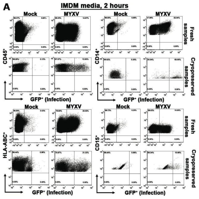
Methods: Bone marrow (BM) and mobilized peripheral blood stem cells (mPBSCs) from patients with hematologic malignancies were treated with MYXV at various time, temperature and incubation media conditions. Treated BM cells from healthy normal donors were evaluated using flow cytometry for MYXV infection, long-term culture-initiating cell (LTC-IC) assay and colony-forming cell (CFC) assay.

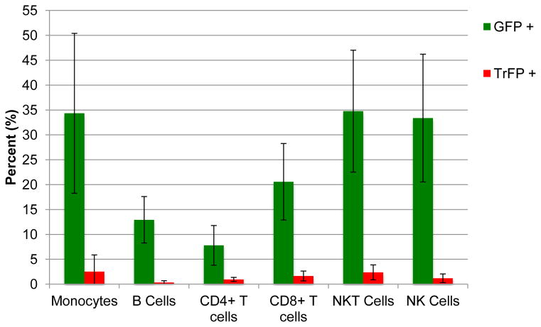
Results: MYXV initiated infection in up to 45% of antigen-presenting monocytes, B cells and natural killer cells; however, these infections were uniformly aborted in >95% of all cells. Fresh graft sources showed higher levels of MYXV infection initiation than cryopreserved specimens, but in all cases less than 10% of CD34(+) cells could be infected after ex vivo MYXV treatment. MYXV did not impair LTC-IC colony numbers compared with mock treatment. CFC colony types and numbers were also not impaired by MYXV treatment. MYXV incubation time, temperature or culture media did not significantly change the percentage of infected cells, LTC-IC colony formation or CFC colony formation.

Conclusions: Human hematopoietic cells are non-permissive for MYXV. Human hematopoietic stem and progenitor cells were not infected and thus unaffected by MYXV ex vivo treatment.

Keywords: bone marrow; mobilized peripheral stem cell blood; myxoma virus; purging; virotherapy.


Figures

Figure 1
Figure 1. MYXV treatment of fresh and cryopreserved bone marrow in various incubation conditions
MYXV treatment of fresh or cryopreserved BM samples from patients with hematological malignancies was performed using a vMyx-GFP construct (MOI of 10) at 37°C. Incubation was carried out in either IMDM, or Plasma-lyte A + 10% ACDA culture media for 2 hours or 24 hours. At the indicated time points, cells were collected and stained with monoclonal antibodies against cell lineages including CD45, HLA-ABC, CD19, CD3, CD34, CD14, CD15, and CD235a. Virus infection of these cell lineages was quantified using flow cytometry. (A and B), Representative 2D-plots showing the percentage of MYXV infection (GFP+) for 2 hours in IMDM and in Plasma-lyte A + 10% ACDA, respectively. (C–J), The percentage of MYXV infection (GFP+) of fresh vs. cryopreserved BM samples, for 2 hours or 24 hours, in IMDM or in Plasma-lyte A + 10% ACDA are shown. Values are mean ± SEM of at least four independent experiments with three independent specimens.
Figure 2
Figure 2. MYXV treatment of fresh and cryopreserved mobilized peripheral blood stem cells in various incubation conditions
MYXV treatment of fresh or cryopreserved mobilized peripheral blood stem cell samples (mPBSCs) from patients with hematological malignancies was performed using a vMyx-GFP construct (MOI of 10) at 37°C. Incubation was carried out in either IMDM, or Plasma-lyte A + 10% ACDA culture media for 2 hours or 24 hours. At the indicated time points, cells were collected and stained with monoclonal antibodies against cell lineages including CD45, HLA-ABC, CD19, CD3, CD34, CD14, CD15, and CD235a. Virus infection of these cell lineages was quantified using flow cytometry. (A and B), Representative 2D-plots showing the percentage of MYXV infection (GFP+) for 2 hours in IMDM and in Plasma-lyte A + 10% ACDA, respectively. (C–J), The percentage of MYXV infection (GFP+) of fresh vs. cryopreserved BM samples, for 2 hours or 24 hours, in IMDM or in Plasma-lyte A + 10% ACDA are shown. Values are mean ± SEM of at least four independent experiments with three independent specimens.
Figure 3
Figure 3. Human peripheral blood mononuclear cells are nonpermissive to MYXV
To determine whether MYXV is permissive to the cell subsets, CD14+ (monocytes), CD3+, CD4+ and CD8+ (T cells), CD19+ (B cells) and CD56+ (NK cells), PBMCs were isolated from healthy donors and then incubated with vMyx-GFP/TrFR (MOI =10) at 4°C for 1 hour. Unbound virus was washed and cells were incubated at 37°C for 24 hours. Next cells stained with the respective fluorochrome-conjugated antibodies, and the proportion of cells expressing GFP+ or TrFP+ in each hematopoietic cell subset was quantified using flow cytometry. Although a high proportion of monocytes expressed GFP+, in all hematopoietic subsets, greater than 95% of cells lacked TrFP expression, indicating that even though early MYXV infection may have initiated in some healthy human hematopoietic cells, the MYXV infection was aborted and the cells were non-permissive to the virus.
Figure 4
Figure 4. Effect of MYXV treatment on LTC-IC from human bone marrow
(A) Micrographs of LITC-ICs after treatment with MYXV. (B) Fresh or (C) cryopreserved BM was treated with vMyx-GFP (MOI of 10) in various culture media including IMDM, Plasma-lyte A + 10% ACDA, heparin, and M199. The hematopoietic stem cell content was quantified by long-term culture-initiating cell (LTC-IC) assay. Data shown correspond to the mean ± SEM of at least three independent specimens, each one performed in duplicate.
Figure 5
Figure 5. Effect of MYXV treatment on CFC from human bone marrow
(A) Micrographs of CFC after treatment with MYXV. (B) Colony formations observed after 14 days according to various MYXV incubation media conditions. (C) CFC colony formations observed after 14 days according to various MYXV incubation temperature conditions. Data shown are the mean ± SEM of at least three independent specimens, each one performed in duplicate.

Similar articles

Cited by

References

    1. Gribben JG, Nadler LM. Bone marrow purging for autologous bone marrow transplantation. Leuk Lymphoma. 1993;11(Suppl 2):141–148. - PubMed
    1. Kvalheim G. Purging of autografts: methods and clinical significance. Ann Med. 1996;28(2):167–173. - PubMed
    1. Rahman MM, Madlambayan GJ, Cogle CR, McFadden G. Oncolytic viral purging of leukemic hematopoietic stem and progenitor cells with Myxoma virus. Cytokine Growth Factor Rev. 2010;21(2–3):169–175. - PMC - PubMed
    1. Yang H, Eaves C, de Lima MLM, et al. A novel triple purge strategy for eliminating chronic myelogenous leukemia (CML) cells from autografts. Bone Marrow Transplant. 2006;37(6):575–582. - PubMed
    1. Vellenga E, van Putten W, Ossenkoppele GJ, et al. Autologous peripheral blood stem cell transplantation for acute myeloid leukemia. Blood. 2011;118(23):6037–6042. - PubMed

Publication types

MeSH terms