

The effect of bisphosphonates on the endothelial differentiation of mesenchymal stem cells
- PMID: 26857282
- PMCID: PMC4746673
- DOI: 10.1038/srep20580
The effect of bisphosphonates on the endothelial differentiation of mesenchymal stem cells
Abstract
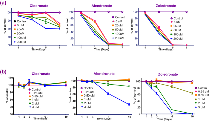
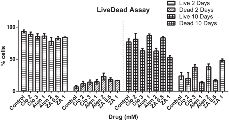
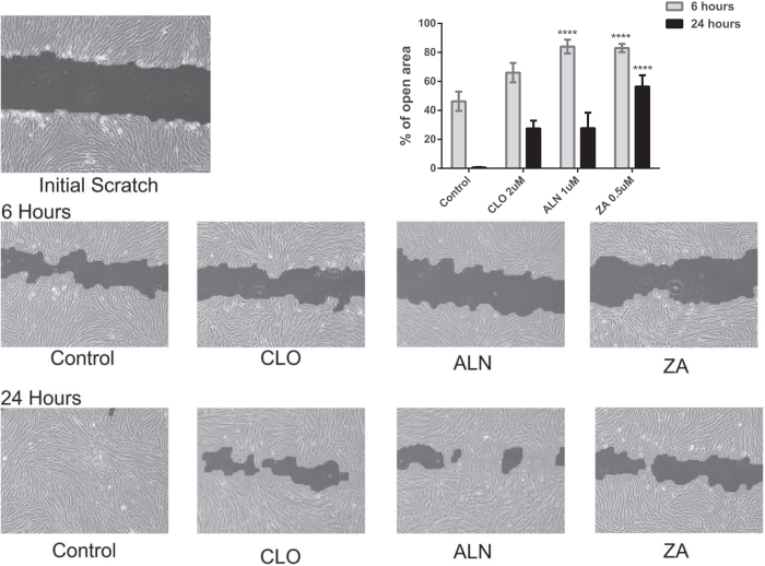
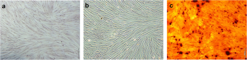
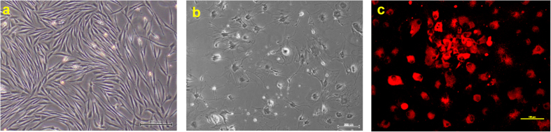
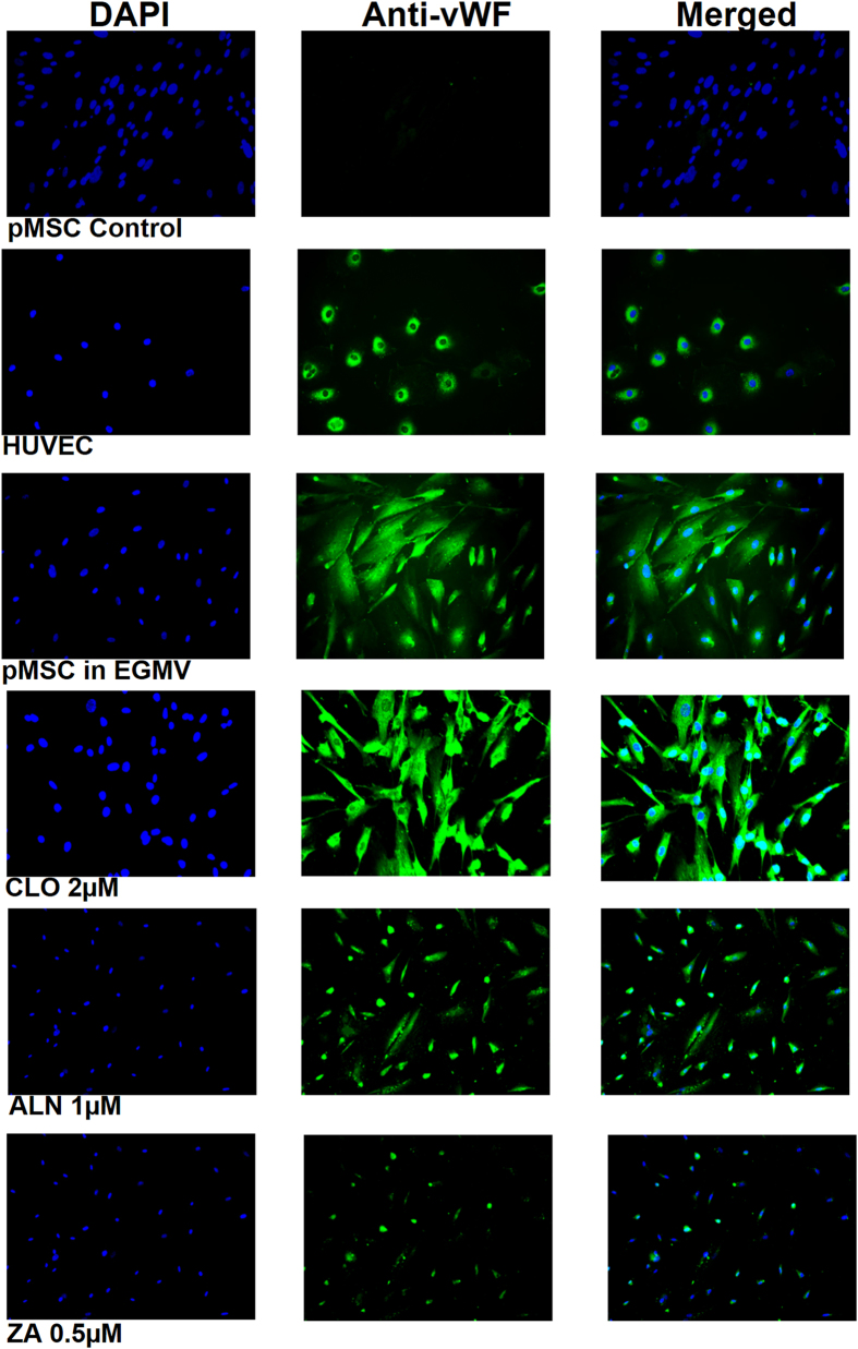
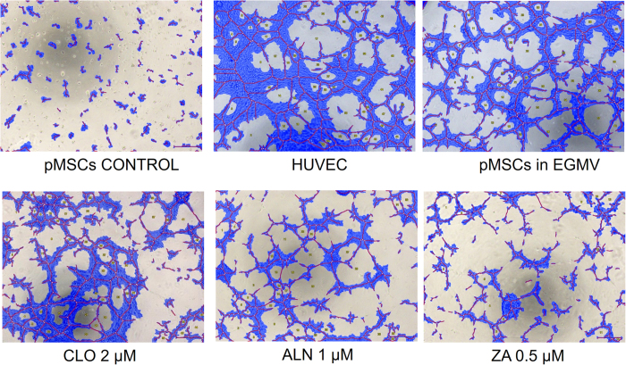
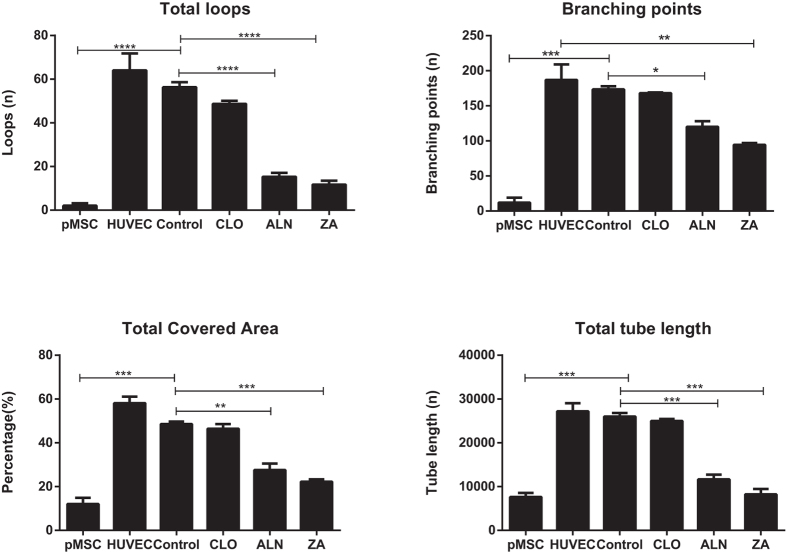
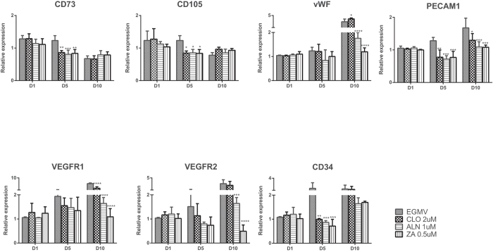
The contribution of the local stem cell niche to providing an adequate vascular framework during healing cannot be overemphasized. Bisphosphonates (BPs) are known to have a direct effect on the local vasculature, but their effect on progenitor cell differentiation is unknown. This in vitro study evaluated the effect(s) of various BPs on the differentiation of human placental mesenchymal stem cells (pMSCs) along the endothelial lineage and their subsequent functional and morphogenic capabilities. pMSC multipotency was confirmed by successful differentiation into cells of both the osteogenic and endothelial lineages, as demonstrated by positive Alizarin Red S staining and Ac-LDL uptake. pMSC differentiation in the presence of non-cytotoxic BP concentrations showed that nitrogen containing BPs had a significant inhibitory effect on cell migration and endothelial marker gene expression, as well as compromised endothelial differentiation as demonstrated using von Willebrand factor immunofluorescence staining and tube formation assay. This in vitro study demonstrated that at non-cytotoxic levels, nitrogen-containing BPs inhibit differentiation of pMSCs into cells of an endothelial lineage and affect the downstream functional capability of these cells supporting a multi-modal effect of BPs on angiogenesis as pathogenic mechanism contributing to bone healing disorders such as bisphosphonate related osteonecrosis of the jaws (BRONJ).
Figures
Similar articles
-
Osteonecrosis of the jaw: effect of bisphosphonate type, local concentration, and acidic milieu on the pathomechanism.J Oral Maxillofac Surg. 2010 Nov;68(11):2837-45. doi: 10.1016/j.joms.2010.07.017. J Oral Maxillofac Surg. 2010. PMID: 20971371
-
Endothelial cells direct human mesenchymal stem cells for osteo- and chondro-lineage differentiation through endothelin-1 and AKT signaling.Stem Cell Res Ther. 2015 May 1;6(1):88. doi: 10.1186/s13287-015-0065-6. Stem Cell Res Ther. 2015. PMID: 25998005 Free PMC article.
-
Simultaneous cultivation of human endothelial-like differentiated precursor cells and human marrow stromal cells on beta-tricalcium phosphate.Tissue Eng Part C Methods. 2009 Dec;15(4):551-60. doi: 10.1089/ten.TEC.2008.0385. Tissue Eng Part C Methods. 2009. PMID: 19199563
-
Osteogenic differentiation of rat mesenchymal stem cells from adipose tissue in comparison with bone marrow mesenchymal stem cells: melatonin as a differentiation factor.Iran Biomed J. 2008 Jul;12(3):133-41. Iran Biomed J. 2008. PMID: 18762816
-
Endothelial cells stimulate osteogenic differentiation of mesenchymal stem cells on calcium phosphate scaffolds.J Tissue Eng Regen Med. 2014 Oct;8(10):831-40. doi: 10.1002/term.1590. Epub 2012 Oct 5. J Tissue Eng Regen Med. 2014. PMID: 23038605
Cited by
-
Current Understanding of the Pathophysiology of Osteonecrosis of the Jaw.Curr Osteoporos Rep. 2018 Oct;16(5):584-595. doi: 10.1007/s11914-018-0474-4. Curr Osteoporos Rep. 2018. PMID: 30155844 Review.
-
Effect of anti-angiogenesis induced by chemotherapeutic monotherapy, chemotherapeutic/bisphosphonate combination therapy and anti-VEGFA mAb therapy on tooth extraction socket healing in mice.J Bone Miner Metab. 2018 Sep;36(5):547-559. doi: 10.1007/s00774-017-0872-1. Epub 2017 Oct 17. J Bone Miner Metab. 2018. PMID: 29043461
-
Angiogenesis in the Development of Medication-Related Osteonecrosis of the Jaws: An Overview.Dent J (Basel). 2016 Dec 26;5(1):2. doi: 10.3390/dj5010002. Dent J (Basel). 2016. PMID: 29563407 Free PMC article. Review.
-
Effect of combined treatment with bisphosphonate and vitamin D on atherosclerosis in patients with systemic lupus erythematosus: a propensity score-based analysis.Arthritis Res Ther. 2018 Apr 17;20(1):72. doi: 10.1186/s13075-018-1589-9. Arthritis Res Ther. 2018. PMID: 29665863 Free PMC article.
-
Skeletal Site-specific Effects of Zoledronate on in vivo Bone Remodeling and in vitro BMSCs Osteogenic Activity.Sci Rep. 2017 Jan 31;7:36129. doi: 10.1038/srep36129. Sci Rep. 2017. PMID: 28139685 Free PMC article.
References
-
- Harada S. & Rodan G. A. Control of osteoblast function and regulation of bone mass. Nature 423, 349–355 (2003). - PubMed
-
- Parfitt A. M. Skeletal Heterogeneity and the Purposes of Bone Remodeling: Implications for the Understanding of Osteoporosis. in Osteoporosis 71–89 (Elsevier BV, 2008).
-
- Shi S. & Gronthos S. Perivascular niche of postnatal mesenchymal stem cells in human bone marrow and dental pulp. J Bone Miner Res 18, 696–704 (2003). - PubMed
-
- Berenson J. R. Antitumor effects of bisphosphonates: from the laboratory to the clinic. Curr Opin Support Palliat Care 5, 233–240 (2011). - PubMed
-
- Terpos E., Dimopoulos M. A. & Berenson J. Established role of bisphosphonate therapy for prevention of skeletal complications from myeloma bone disease. Crit Rev Oncol Hematol 77 Suppl 1, S13–23 (2011). - PubMed
Publication types
MeSH terms
Substances
LinkOut - more resources
Full Text Sources
Other Literature Sources
