

Clinicopathologic, Immunohistochemical, and Ultrastructural Findings of a Fatal Case of Middle East Respiratory Syndrome Coronavirus Infection in the United Arab Emirates, April 2014
- PMID: 26857507
- PMCID: PMC7093852
- DOI: 10.1016/j.ajpath.2015.10.024
Clinicopathologic, Immunohistochemical, and Ultrastructural Findings of a Fatal Case of Middle East Respiratory Syndrome Coronavirus Infection in the United Arab Emirates, April 2014
Abstract
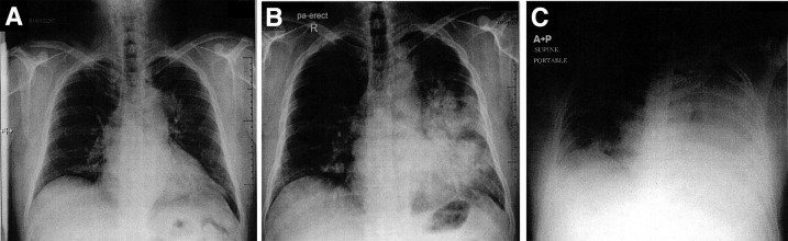
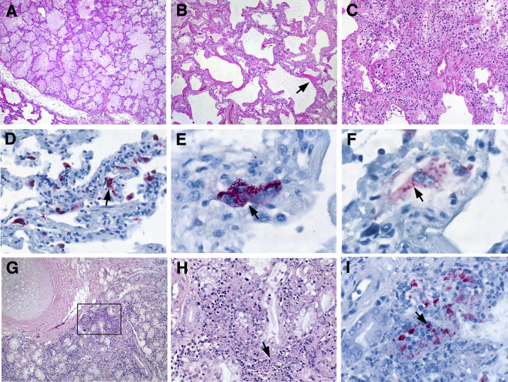
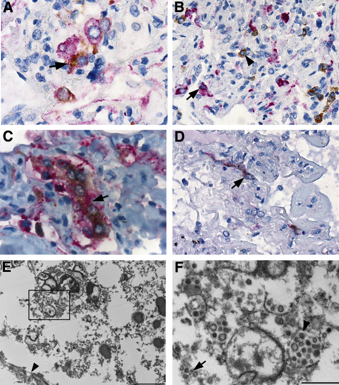
Middle East respiratory syndrome coronavirus (MERS-CoV) infection causes an acute respiratory illness and is associated with a high case fatality rate; however, the pathogenesis of severe and fatal MERS-CoV infection is unknown. We describe the histopathologic, immunohistochemical, and ultrastructural findings from the first autopsy performed on a fatal case of MERS-CoV in the world, which was related to a hospital outbreak in the United Arab Emirates in April 2014. The main histopathologic finding in the lungs was diffuse alveolar damage. Evidence of chronic disease, including severe peripheral vascular disease, patchy cardiac fibrosis, and hepatic steatosis, was noted in the other organs. Double staining immunoassays that used anti-MERS-CoV antibodies paired with immunohistochemistry for cytokeratin and surfactant identified pneumocytes and epithelial syncytial cells as important targets of MERS-CoV antigen; double immunostaining with dipeptidyl peptidase 4 showed colocalization in scattered pneumocytes and syncytial cells. No evidence of extrapulmonary MERS-CoV antigens were detected, including the kidney. These results provide critical insights into the pathogenesis of MERS-CoV in humans.
Copyright © 2016 American Society for Investigative Pathology. Published by Elsevier Inc. All rights reserved.
Figures
Comment in
-
Value of Autopsy Emphasized in the Case Report of a Single Patient with Middle East Respiratory Syndrome.Am J Pathol. 2016 Mar;186(3):507-10. doi: 10.1016/j.ajpath.2015.11.001. Epub 2015 Dec 24. Am J Pathol. 2016. PMID: 26724388 Free PMC article.
-
[The value of one autopsy].Medicina (B Aires). 2017;77(3):245-246. Medicina (B Aires). 2017. PMID: 28643687 Spanish. No abstract available.
References
-
- Zaki A.M., van Boheemen S., Bestebroer T.M., Osterhaus A.D., Fouchier R.A. Isolation of a novel coronavirus from a man with pneumonia in Saudi Arabia. N Engl J Med. 2012;367:1814–1820. - PubMed
-
- Azhar E.I., El-Kafrawy S.A., Farraj S.A., Hassan A.M., Al-Saeed M.S., Hashem A.M., Madani T.A. Evidence for camel-to-human transmission of MERS coronavirus. N Engl J Med. 2014;370:2499–2505. - PubMed
-
- Haagmans B.L., Al Dhahiry S.H., Reusken C.B., Raj V.S., Galiano M., Myers R., Godeke G.J., Jonges M., Farag E., Diab A., Ghobashy H., Alhajri F., Al-Thani M., Al-Marri S.A., Al Romaihi H.E., Al Khal A., Bermingham A., Osterhaus A.D., AlHajri M.M., Koopmans M.P. Middle East respiratory syndrome coronavirus in dromedary camels: an outbreak investigation. Lancet Infect Dis. 2014;14:140–145. - PMC - PubMed
-
- Assiri A., McGeer A., Perl T.M., Price C.S., Al Rabeeah A.A., Cummings D.A., Alabdullatif Z.N., Assad M., Almulhim A., Makhdoom H., Madani H., Alhakeem R., Al-Tawfiq J.A., Cotten M., Watson S.J., Kellam P., Zumla A.I., Memish Z.A., KSA MERS-CoV Investigation Team Hospital outbreak of Middle East respiratory syndrome coronavirus. N Engl J Med. 2013;369:407–416. - PMC - PubMed
-
- Al-Abdallat M.M., Payne D.C., Alqasrawi S., Rha B., Tohme R.A., Abedi G.R., Al Nsour M., Iblan I., Jarour N., Farag N.H., Haddadin A., Al-Sanouri T., Tamin A., Harcourt J.L., Kuhar D.T., Swerdlow D.L., Erdman D.D., Pallansch M.A., Haynes L.M., Gerber S.I., Jordan MERS-CoV Investigation Team Hospital-associated outbreak of Middle East respiratory syndrome coronavirus: a serologic, epidemiologic, and clinical description. Clin Infect Dis. 2014;59:1225–1233. - PMC - PubMed
Publication types
MeSH terms
Substances
LinkOut - more resources
Full Text Sources
Other Literature Sources
Miscellaneous
