. 2016 Mar 24;84(4):1214-1225.
doi: 10.1128/IAI.01462-15. Print 2016 Apr.

XerC Contributes to Diverse Forms of Staphylococcus aureus Infection via agr-Dependent and agr-Independent Pathways

Affiliations

XerC Contributes to Diverse Forms of Staphylococcus aureus Infection via agr-Dependent and agr-Independent Pathways

Danielle N Atwood et al. Infect Immun. .

Abstract

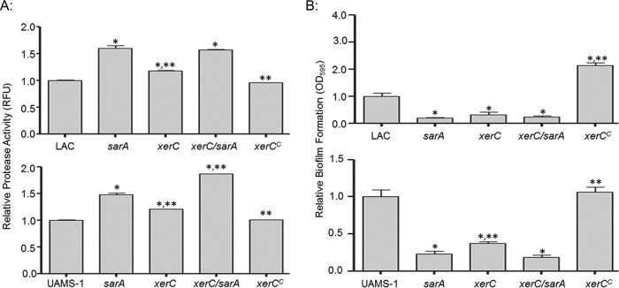
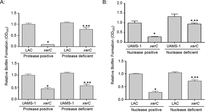
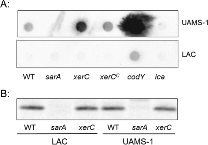
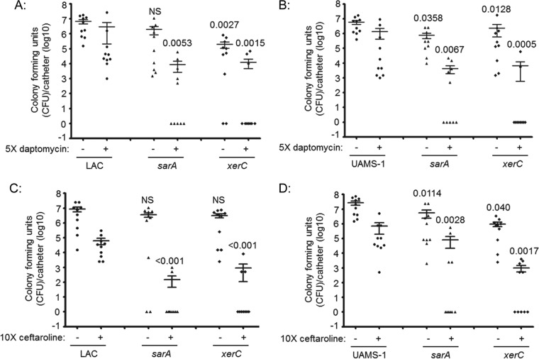
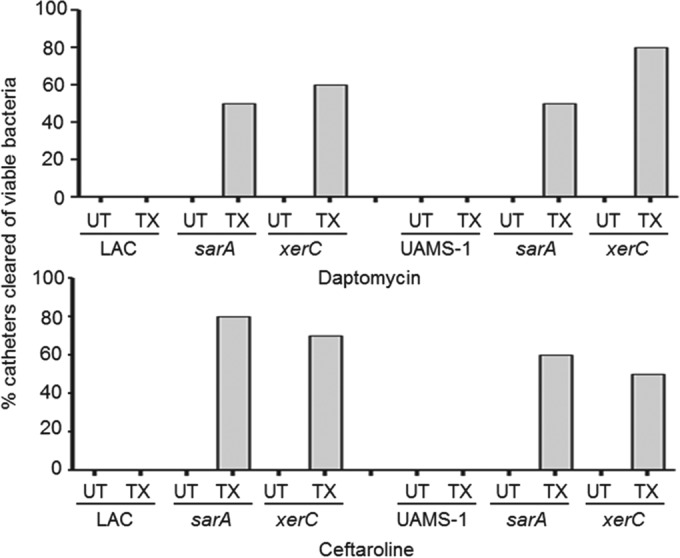
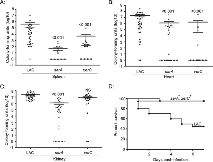
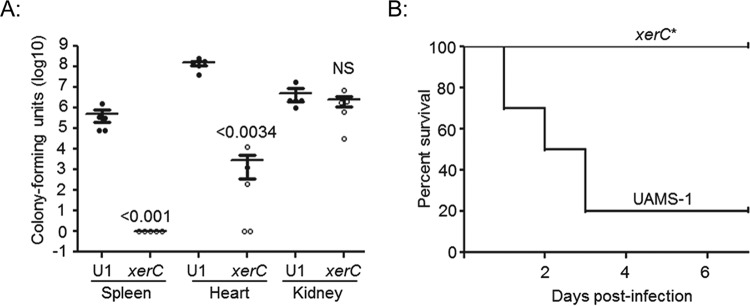
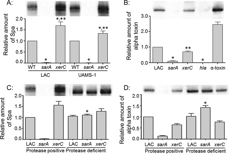
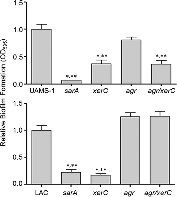
We demonstrate that mutation of xerC, which reportedly encodes a homologue of an Escherichia coli recombinase, limits biofilm formation in the methicillin-resistant Staphylococcus aureus strain LAC and the methicillin-sensitive strain UAMS-1. This was not due to the decreased production of the polysaccharide intracellular adhesin (PIA) in either strain because the amount of PIA was increased in a UAMS-1xerC mutant and undetectable in both LAC and its isogenic xerC mutant. Mutation of xerC also resulted in the increased production of extracellular proteases and nucleases in both LAC and UAMS-1, and limiting the production of either class of enzymes increased biofilm formation in the isogenic xerC mutants. More importantly, the limited capacity to form a biofilm was correlated with increased antibiotic susceptibility in both strains in the context of an established biofilm in vivo. Mutation of xerC also attenuated virulence in a murine bacteremia model, as assessed on the basis of the bacterial loads in internal organs and overall lethality. It also resulted in the decreased accumulation of alpha toxin and the increased accumulation of protein A. These findings suggest that xerC may impact the functional status of agr. This was confirmed by demonstrating the reduced accumulation of RNAIII and AgrA in LAC and UAMS-1xerC mutants. However, this cannot account for the biofilm-deficient phenotype of xerC mutants because mutation of agr did not limit biofilm formation in either strain. These results demonstrate that xerC contributes to biofilm-associated infections and acute bacteremia and that this is likely due to agr-independent and -dependent pathways, respectively.


Figures

FIG 1
FIG 1
Impact of xerC on protease production and biofilm formation. (A) Protease production in LAC (top), UAMS-1 (bottom), their isogenic derivatives with mutations in sarA and/or xerC, and their genetically complemented xerC mutants (xerCC) was assessed using a fluorescence resonance energy transfer-based assay. (B) Biofilm formation in LAC (top), UAMS-1 (bottom), isogenic derivatives with mutations in sarA and/or xerC, and complemented xerC mutants (xerCC) was assessed using a microtiter plate assay. Permutation-based ANOVA models were used to perform these analyses. *, a significant difference relative to the result for the isogenic parent strain; **, a significant difference by comparison to the result for the isogenic sarA mutant. RFU, relative fluorescence units.
FIG 2
FIG 2
Relative biofilm formation in xerC mutants as a function of protease and nuclease production. (A) Biofilm formation in LAC (top), UAMS-1 (bottom), their respective xerC mutants, and their isogenic xerC mutants unable to produce all (LAC) or most (UAMS-1) S. aureus extracellular proteases was assessed using a microtiter plate assay. (B) Biofilm formation in UAMS-1, LAC, their isogenic xerC mutants, and their nuclease-deficient xerC mutants unable to produce the primary S. aureus extracellular nuclease (Nuc1) was assessed. Permutation-based ANOVA models were used to perform these analyses. *, a statistically significant difference between the xerC mutant and its isogenic parent strain with the equivalent capacity to produce extracellular proteases (A) or nucleases (B); **, a statistically significant difference between the protease or nuclease-deficient xerC mutant and the isogenic protease or nuclease-positive xerC strain.
FIG 3
FIG 3
Impact of xerC on PIA and SarA production. (A) PIA production in UAMS-1, LAC, their isogenic sarA, xerC, codY, and ica mutants, and their genetically complemented xerC mutants (xerCC) was assessed by Western dot blotting. The ica and codY mutants were included as negative and positive controls, respectively, on the basis of the fact that ica encodes the enzymes necessary for PIA production, while mutation of codY was previously shown to result in increased PIA production in UAMS-1 (17). (B) The abundance of SarA was assessed by conventional Western blotting. The results shown are representative of those from at least three biological replicates. WT, wild type.
FIG 4
FIG 4
Impact of xerC on biofilm formation and antibiotic susceptibility in vivo. Catheters were implanted into the flanks of mice prior to colonization with LAC (A and C), UAMS-1 (B and D), or their isogenic sarA or xerC mutants. After 24 h, the lumen of each catheter was injected with sterile PBS (−) or the antibiotic (+) daptomycin (A and B) or ceftaroline (C and D). This was continued daily for 5 days. At 24 h after the last injection, the catheters were recovered and processed to assess the number of CFU remaining per catheter. The horizontal line within each group indicates the mean for that experimental group. Numbers above each group are permutation-based unpaired t-test P values for that group compared to the result for the parent strain with or without antibiotic exposure as appropriate. NS, not significant.
FIG 5
FIG 5
Percentage of catheters cleared by antibiotic exposure. Results illustrate the percentage of catheters (n = 10) colonized with LAC, UAMS-1, or their isogenic xerC and sarA mutants that were cleared of viable bacteria with exposure (treated [TX]) and without exposure (untreated [UT]) to the indicated antibiotic.
FIG 6
FIG 6
Virulence of LAC xerC mutants in hematogenous infection. Mice were infected by tail vein injection with the indicated strains. Tissues were harvested either upon compassionate euthanasia or after 6 days. The results shown are the colony counts obtained from each tissue in individual mice, with the horizontal line within each group indicating the mean within that experimental group (A to C). Permutation-based ANOVA models were used to analyze these data. The numbers above each group are P values for that group compared to the result for the isogenic parent strain. NS, not significant. (D) Kaplan-Meier survival curves for mice hematogenously infected with LAC and its isogenic xerC or sarA mutant. Log rank tests were used to compare the results for the mutant and parent strains. *, statistically significant difference by comparison to the result for the parent strain.
FIG 7
FIG 7
Virulence of UAMS-1 xerC mutants in hematogenous infection. Mice were infected by tail vein injection with UAMS-1 (U1) or its isogenic xerC mutant. Tissues were harvested after compassionate euthanasia or at 6 days after infection. (A) The results shown are the colony counts obtained from each tissue in individual mice, with the horizontal line within each group indicating the mean within that experimental group. Permutation-based unpaired t tests were used to compare the result for the isogenic xerC mutant to that for the parent strain. The numbers above each xerC mutant are P values for that group compared to the result for the parent strain. NS, not significant. (B) Kaplan-Meier survival curves for mice hematogenously infected with UAMS-1 and its isogenic xerC mutant. *, a statistically significant difference by the log-rank test when the result for the isogenic xerC mutant is compared to that for the parent strain.
FIG 8
FIG 8
Abundance of protein A and alpha toxin in xerC mutants. (A) The relative abundance of protein A (Spa), as assessed by measuring the amount of extracellular Spa (eSpa) in clarified supernatants, was assessed in LAC, UAMS-1, and the indicated mutants by Western blotting. (B) The relative amounts of alpha toxin in LAC and the indicated mutants were assessed by Western blotting. An alpha toxin mutant (hla) and purified alpha toxin were included as controls. UAMS-1 does not produce alpha toxin and thus was not included in this experiment. *, a statistically significant difference by comparison to the results for the isogenic parent strain; **, a statistically significant difference by comparison to the results for the isogenic sarA mutant. (C and D) The relative amounts of eSpa (C) and alpha toxin (D) in LAC, its sarA and xerC mutants, and protease-deficient derivatives of each of these strains were assessed by Western blotting. *, a statistically significant difference between the results for the protease-deficient derivative and the isogenic protease-positive strain. Permutation-based ANOVA models were used to perform these analyses.
FIG 9
FIG 9
Impact of xerC on agr. (A) The abundance of AgrA in LAC, UAMS-1, their isogenic sarA, xerC and agr mutants, and their genetically complemented xerC mutants (xerCC) was assessed by Western blotting. (B) The abundance of RNAIII in the indicated strains was assessed by qRT-PCR. *, a statistically significant decrease relative to the amount for the isogenic parent strain. Permutation-based ANOVA models were used to perform these analyses.
FIG 10
FIG 10
Relative impact of agr and xerC on biofilm formation. Biofilm formation was assessed in UAMS-1 (top), LAC (bottom), and their isogenic sarA, agr, xerC, and agr/xerC mutants. *, a statistically significant difference relative to the result for the isogenic parent strain; **, a statistically significant difference by comparison to the result for the isogenic agr mutant. Permutation-based ANOVA models were used to perform these analyses.

Similar articles

Cited by

References

    1. Laabei M, Uhlemann AC, Lowy FD, Austin ED, Yokoyama M, Ouadi K, Feil E, Thorpe HA, Williams B, Perkins M, Peacock SJ, Clarke SR, Dordel J, Holden M, Votintseva AA, Bowden R, Crook DW, Young BC, Wilson DJ, Recker M, Massey RC. 2015. Evolutionary trade-offs underlie the multi-faceted virulence of Staphylococcus aureus. PLoS Biol 13:e1002229. doi:10.1371/journal.pbio.1002229. - DOI - PMC - PubMed
    1. Lowy FD. 1998. Staphylococcus aureus infections. N Engl J Med 339:520–532. doi:10.1056/NEJM199808203390806. - DOI - PubMed
    1. Beenken KE, Mrak LN, Griffin LM, Zielinska AK, Shaw LN, Rice KC, Horswill AR, Bayles KW, Smeltzer MS. 2010. Epistatic relationships between sarA and agr in Staphylococcus aureus biofilm formation. PLoS One 5:e10790. doi:10.1371/journal.pone.0010790. - DOI - PMC - PubMed
    1. Boles BR, Horswill AR. 2008. Agr-mediated dispersal of Staphylococcus aureus biofilms. PLoS Pathog 4:e1000052. doi:10.1371/journal.ppat.1000052. - DOI - PMC - PubMed
    1. Bassetti M, Merelli M, Temperoni C, Astilean A. 2013. New antibiotics for bad bugs: where are we? Ann Clin Microbiol Antimicrob 12:22. doi:10.1186/1476-0711-12-22. - DOI - PMC - PubMed

Publication types

MeSH terms

LinkOut - more resources