. 2016 Mar 24;84(4):1194-1204.
doi: 10.1128/IAI.01390-15. Print 2016 Apr.

Porphyromonas gingivalis Outer Membrane Vesicles Induce Selective Tumor Necrosis Factor Tolerance in a Toll-Like Receptor 4- and mTOR-Dependent Manner

Affiliations

Porphyromonas gingivalis Outer Membrane Vesicles Induce Selective Tumor Necrosis Factor Tolerance in a Toll-Like Receptor 4- and mTOR-Dependent Manner

Tobias Waller et al. Infect Immun. .

Abstract

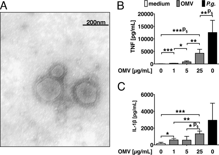
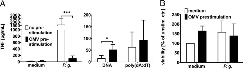
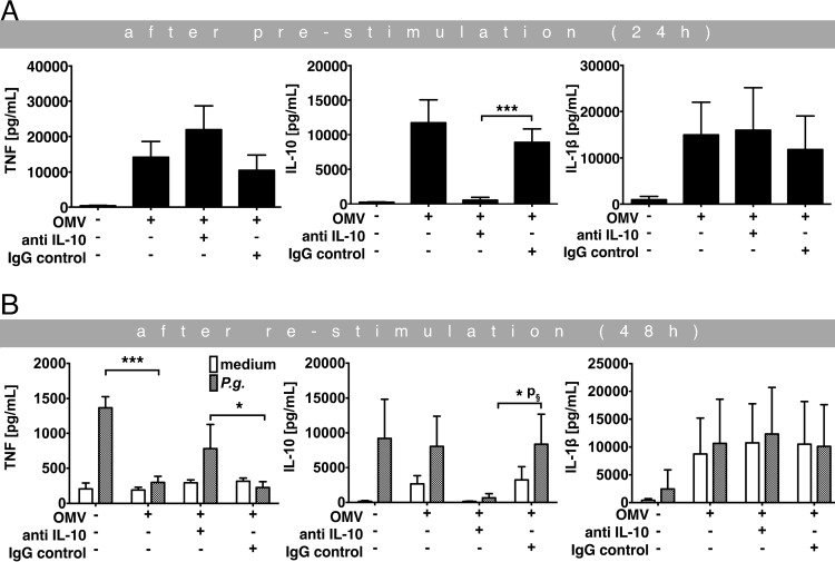
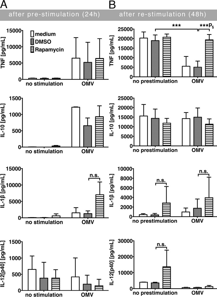
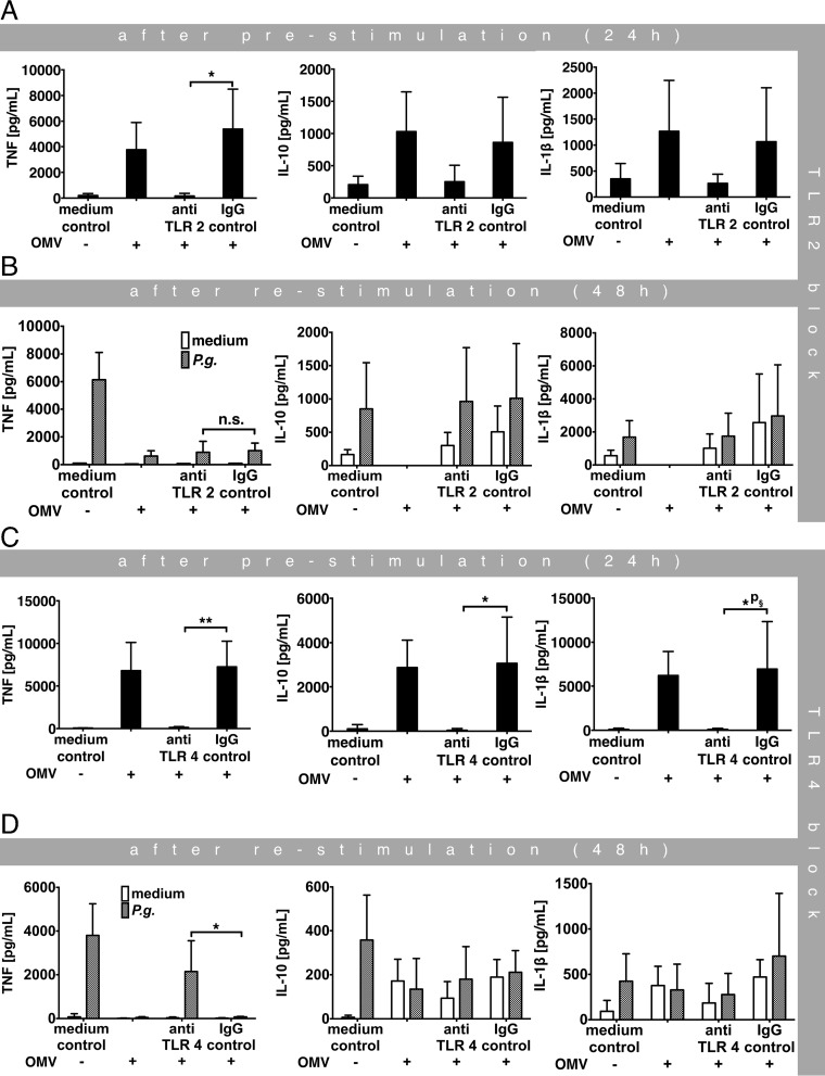
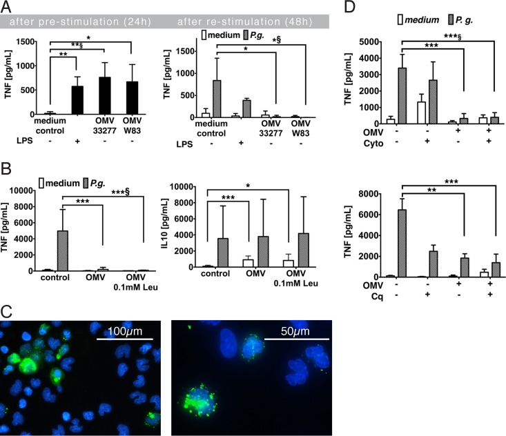
Porphyromonas gingivalis is an important member of the anaerobic oral flora. Its presence fosters growth of periodontal biofilm and development of periodontitis. In this study, we demonstrated that lipophilic outer membrane vesicles (OMV) shed from P. gingivalis promote monocyte unresponsiveness to live P. gingivalis but retain reactivity to stimulation with bacterial DNA isolated from P. gingivalis or AIM2 ligand poly(dA·dT). OMV-mediated tolerance of P. gingivalis is characterized by selective abrogation of tumor necrosis factor (TNF). Neutralization of interleukin-10 (IL-10) during OMV challenge partially restores monocyte responsiveness toP. gingivalis; full reactivity toP. gingivalis can be restored by inhibition of mTOR signaling, which we previously identified as the major signaling pathway promoting Toll-like receptor 2 and Toll-like receptor 4 (TLR2/4)-mediated tolerance in monocytes. However, despite previous reports emphasizing a central role of TLR2 in innate immune recognition of P. gingivalis, our current findings highlight a selective role of TLR4 in the promotion of OMV-mediated TNF tolerance: only blockade of TLR4-and not of TLR2-restores responsiveness toP. gingivalis Of further note, OMV-mediated tolerance is preserved in the presence of cytochalasin B and chloroquine, indicating that triggering of surface TLR4 is sufficient for this effect. Taking the results together, we propose that P. gingivalis OMV contribute to local immune evasion of P. gingivalis by hampering the host response.


Figures

FIG 1
FIG 1
Secretion of proinflammatory cytokines in response to P. gingivalis OMV. (A) Trans-electron microscopy of the purified OMV. The image shows a negative stain with 2% methylamine tungstate and is representative of the results of n = 6 experiments. (B and C) Human CD14+ monocytes were stimulated with OMV at the indicated concentrations or with live P. gingivalis (P.g.). Cellular supernatants were harvested after 24 h and cytokines quantified by ELISA. (B) TNF. (C) IL-1β. The graphs summarize the results obtained from n = 6 independent donors (n = 3 independent experiments). ***, P = 0.0002; ***p§ = 0.0008; *, P = 0.0281; **, P = 0.0031; **p§ = 0.0027 (B); *, P = 0.0119; ***, P = 0.0003; **, P = 0.0051; *p§ = 0.0217 (C).
FIG 2
FIG 2
Effects of OMV preexposure on TNF secretion in response to secondary stimulation with live P. gingivalis. (A) Human monocytes were prestimulated or not prestimulated with OMV for 24 h; after washing, cells were left unstimulated (medium) or restimulated with live P. gingivalis, P. gingivalis DNA, or poly(dA·dT) for another 24 h. TNF secretion was quantified in the monocyte supernatants. Results are shown as mean values ± standard deviations (SD) of data from 12 donors (n = 6 independent experiments). ***, P < 0.0001; *, P = 0.0329. (B) WST-1 was used to determine viability of OMV-stimulated monocytes after 48 h. Results from n = 12 donors (n = 6 independent experiments) were normalized to the unstimulated control (unstim. ctr.) results, and data are given in percentages (mean values ± SD).
FIG 3
FIG 3
Role of IL-10 in OMV-induced monocyte tolerance. (A and B) Monocytes were left unstimulated or prestimulated with OMV in the presence or absence of anti-IL-10 or the IgG isotype control for 24 h. Cells were subsequently washed and restimulated with or without live P. gingivalis for an additional 24 h. TNF, IL-10, and IL-1β levels were measured in the cellular supernatants obtained after prestimulation (24 h) (A) and after secondary challenge (48 h) (B). The results are shown as mean values ± SD of data from n = 4 donors (n = 2 independent experiments). ***, P = 0.0002 (A); *, P = 0.02083; *p§ = 0.0121; ***, P < 0.0001 (B).
FIG 4
FIG 4
Role of mTOR in OMV-induced TNF tolerance. (A and B) CD14+ monocytes were preincubated with or without OMV in the presence or absence of rapamycin DMSO as a solvent control for 24 h. Restimulation after washing of cells was performed with or without live P. gingivalis. Only data from conditions that included P. gingivalis are shown in the diagrams. Concentrations of TNF, IL-10, IL-1β, and IL-12p40 were quantified in the supernatants collected after prestimulation (24 h) (A) and restimulation (48 h) (B). Data are provided as mean values ± SD of results from n = 6 donors (n = 3 independent experiments). ***, P = 0.00071; ***p§ = 0.00019; n.s. = not significant.
FIG 5
FIG 5
Role of TLR2 and TLR4 in OMV-mediated abrogation of the TNF response. (A to D) Human monocytes were prestimulated with OMV or not prestimulated; live P. gingivalis was used for secondary challenge after washing. The experiments were performed in the presence of neutralizing anti-TLR2 (A and B) and anti-TLR4 (C and D) antibodies or of the respective isotype controls. TNF secretion was measured in the supernatants harvested after 24 (A and C) and 48 h (B and D). The diagrams summarize the data obtained as mean values ± SD of the results from n = 4 donors (n = 4 independent experiments). *, P = 0.0154 (A); n.s. = not significant (B); *, P = 0.0281; *p§ = 0.0452; **, P = 0.0032 (C); *, P = 0.0261 (D).
FIG 6
FIG 6
Role of fimbriae, gingipains, and endocytosis in TNF tolerance. (A) Monocytes were left unstimulated or prestimulated with Salmonella Minnesota LPS, P. gingivalis (ATCC 33277) OMV, or OMV of the nonfimbriated P. gingivalis W83 strain for 24 h. Cells were subsequently washed and restimulated with live P. gingivalis (ATCC 33277) for an additional 24 h or not restimulated. TNF, IL-10, and IL-1β levels were measured in the cellular supernatants obtained after prestimulation (24 h) (A) and after secondary challenge (48 h) (B). The results are shown as mean values ± SD of data from n = 4 donors (summary of 2 independent experiments). Left panel: **, P = 0.0023; **p§ = 0.0053; *, P = 0.0143. Right panel: *, P = 0.0487; *p§ = 0.0187. (B) Monocytes were left unstimulated or prestimulated with OMV that had been pretreated with 100 μM leupeptin (Leu). Cells were subsequently washed and restimulated with live P. gingivalis (ATCC 33277) for an additional 24 h or not restimulated. TNF and IL-10 concentrations in the supernatants were measured after secondary challenge (48 h). Results are given as mean values ± SD of data from n = 6 donors (n = 3 independent experiments). Left panel: ***, P = 0.0002; ***p§ = 0.0004. Right panel: ***, P = 0.00042; *, P = 0.02513. (C) Immunofluorescence microscopy was used to visualize cellular binding and internalization of OMV labeled with Vybrant-DIO (green) coincubated with monocytes for 5 h. Nuclei were counterstained with DAPI (blue). The images were acquired with 40-fold magnification (upper panel) and 2× zoom (lower panel) and are representative of the results of n = 2 experiments. Both images were cropped for better visualization. (D) Human monocytes were prestimulated with OMV or not prestimulated in the presence or absence of cytochalasin B (Cyto) (upper panel), chloroquine (Cq) (lower panel), or the DMSO solvent control. Restimulation after washing was performed with or without live P. gingivalis, but only data from conditions that included P. gingivalis are depicted in the graphs. TNF concentrations in the supernatants were measured 24 h after restimulation (48 h). Results are given as mean values ± SD of data from n = 4 independent donors (n = 2 independent experiments). Upper panel: ***, P = 0.00044; ***p§ = 0.00049. Lower panel: **, P = 0.00222; ***, P = 0.00083.

Similar articles

Cited by

References

    1. Shah HN, Gharbia SE. 1995. The biochemical milieu of the host in the selection of anaerobic species in the oral cavity. Clin Infect Dis 20(Suppl 2):S291–S300. - PubMed
    1. Hajishengallis G. 2014. Immunomicrobial pathogenesis of periodontitis: keystones, pathobionts, and host response. Trends Immunol 35:3–11. doi:10.1016/j.it.2013.09.001. - DOI - PMC - PubMed
    1. Hajishengallis G, Darveau RP, Curtis MA. 2012. The keystone-pathogen hypothesis. Nat Rev Microbiol 10:717–725. doi:10.1038/nrmicro2873. - DOI - PMC - PubMed
    1. Ellis TN, Kuehn MJ. 2010. Virulence and immunomodulatory roles of bacterial outer membrane vesicles. Microbiol Mol Biol Rev 74:81–94. doi:10.1128/MMBR.00031-09. - DOI - PMC - PubMed
    1. Haurat MF, Aduse-Opoku J, Rangarajan M, Dorobantu L, Gray MR, Curtis MA, Feldman MF. 2011. Selective sorting of cargo proteins into bacterial membrane vesicles. J Biol Chem 286:1269–1276. doi:10.1074/jbc.M110.185744. - DOI - PMC - PubMed

Publication types

MeSH terms