Case Reports
. 2016 Feb 8:2016:bcr2015209785.
doi: 10.1136/bcr-2015-209785.

Extensive and ulcerated malignant proliferating trichilemmal (pilar) tumour, arising from multiple, large, degenerated trichilemmal (pilar) cysts

Affiliations
Case Reports

Extensive and ulcerated malignant proliferating trichilemmal (pilar) tumour, arising from multiple, large, degenerated trichilemmal (pilar) cysts

Bruno Morgado et al. BMJ Case Rep. .

Abstract

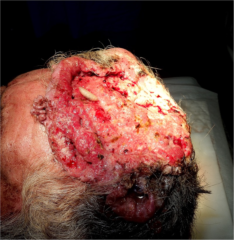
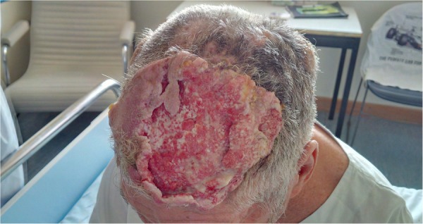
We report a rare case of a 61-year-old homeless man with a 15-year history of multiple trichilemmal cysts that served as a forerunner for the emergence of a malignant proliferating pilar tumour. The patient presented multiple, large, purulent, ulcerated lesions ranging from 10 to 150 mm in diameter, covering most of the scalp, with large areas superimposed by extensive myiasis infestation. The patient presented with no other major clinical findings. A CT scan showed no detectable signs of local or distant metastatic invasion. Initial supportive treatment was implemented. Given the extent of the injury, further surgical excision was considered, which required transfer to a specialised surgical centre. This social case is of educational value, as it can raise clinician awareness about the ability of trichilemmal cysts to undergo malignant transformation. Additionally, it highlights the importance of adequate social assistance structures for patients in need.


Figures

Figure 1
Figure 1
Macroscopic view of the main lesion on admission. The ulcerated area (150×150 mm) covers the frontoparietal aspect of the patient's head. One of the larvae removed from the lesion is also observable.
Figure 2
Figure 2
Macroscopic view of the satellite lesions. Five dome-like formations of various sizes are noticeable on the parietal and occipital epicranium.
Figure 3
Figure 3
Microscopic view of trichilemmal (pilar) cyst wall. The cyst contains amorphous, dense and compact homogenised keratin. There is no granular layer. (H&E ×100).
Figure 4
Figure 4
(A) Microscopic view of the main tumour. Carcinomatous infiltrating component along with cytological atypia and mitosis. (H&E ×100). (B) Microscopic view of the main tumour. Carcinomatous infiltrating component, dissecting eccrine sweat glands. (H&E ×200).
Figure 5
Figure 5
(A–C) CT scan of the head. Five epicranial oval lesions of various sizes, the smallest lesion being 14.5×15 mm, and the largest, 85×80 mm in diameter, with non-pure heterogeneous liquid-air content, forming multiple fluid-air levels. There are no discernable signs of regional tumour invasion.
Figure 6
Figure 6
Macroscopic view of the main lesion 7 days after supportive treatment. The ulcerated area presents signs of improvement, with fibrinous and granulation tissue covering the ulcerated depression.

Similar articles

Cited by

References

    1. Brownstein MH, Arluk DJ. Proliferating trichilemmal cyst: a simulant of squamous cell carcinoma. Cancer 1981;48:1207–14. 10.1002/1097-0142(19810901)48:5<1207::AID-CNCR2820480526>3.0.CO;2-1 - DOI - PubMed
    1. Weedon D, Strutton G, Rubin AI. Weedon's skin pathology. 3rd edn Edinburgh: Churchill Livingstone/Elsevier, 2010:444.
    1. Siddha M, Budrukkar A, Shet T et al. . Malignant pilar tumor of the scalp: a case report and review of literature. J Cancer Res Ther 2007;3:240–3. 10.4103/0973-1482.39001 - DOI - PubMed
    1. Sharma R, Verma P, Yadav P et al. . Proliferating trichilemmal tumor of scalp: benign or malignant, a dilemma. J Cutan Aesthet Surg 2012;5:213–15. 10.4103/0974-2077.101394 - DOI - PMC - PubMed
    1. Jones EW. Proliferating epidermoid cysts. Arch Dermatol 1966;94:11–19. 10.1001/archderm.1966.01600250017002 - DOI - PubMed

Publication types

Supplementary concepts