

Subcortical volumetric abnormalities in bipolar disorder
- PMID: 26857596
- PMCID: PMC5116479
- DOI: 10.1038/mp.2015.227
Subcortical volumetric abnormalities in bipolar disorder
Abstract
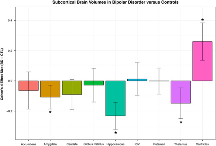
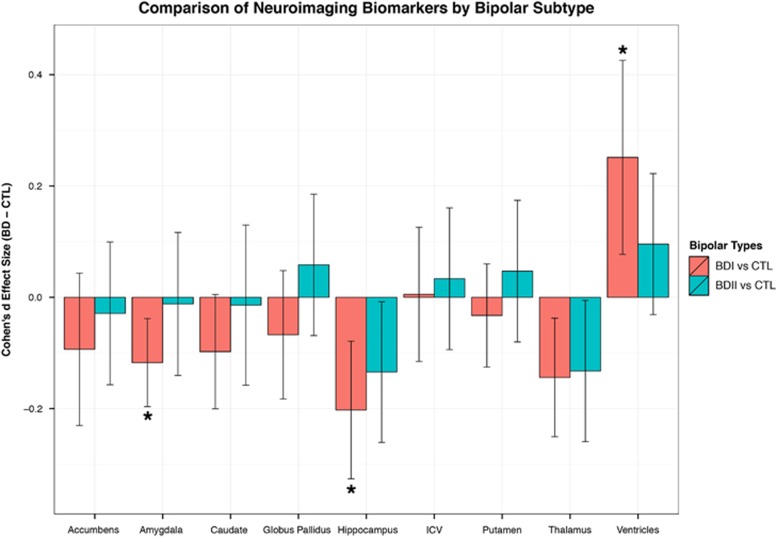
Considerable uncertainty exists about the defining brain changes associated with bipolar disorder (BD). Understanding and quantifying the sources of uncertainty can help generate novel clinical hypotheses about etiology and assist in the development of biomarkers for indexing disease progression and prognosis. Here we were interested in quantifying case-control differences in intracranial volume (ICV) and each of eight subcortical brain measures: nucleus accumbens, amygdala, caudate, hippocampus, globus pallidus, putamen, thalamus, lateral ventricles. In a large study of 1710 BD patients and 2594 healthy controls, we found consistent volumetric reductions in BD patients for mean hippocampus (Cohen's d=-0.232; P=3.50 × 10⁻⁷) and thalamus (d=-0.148; P=4.27 × 10⁻³) and enlarged lateral ventricles (d=-0.260; P=3.93 × 10⁻⁵) in patients. No significant effect of age at illness onset was detected. Stratifying patients based on clinical subtype (BD type I or type II) revealed that BDI patients had significantly larger lateral ventricles and smaller hippocampus and amygdala than controls. However, when comparing BDI and BDII patients directly, we did not detect any significant differences in brain volume. This likely represents similar etiology between BD subtype classifications. Exploratory analyses revealed significantly larger thalamic volumes in patients taking lithium compared with patients not taking lithium. We detected no significant differences between BDII patients and controls in the largest such comparison to date. Findings in this study should be interpreted with caution and with careful consideration of the limitations inherent to meta-analyzed neuroimaging comparisons.
Figures
References
-
- McGuffin P, Rijsdijk F, Andrew M, Sham P, Katz R, Cardno A. The heritability of bipolar affective disorder and the genetic relationship to unipolar depression. Arch Gen Psychiatry 2003; 60: 497–502. - PubMed
-
- McDonald C, Zanelli J, Rabe-Hesketh S, Ellison-Wright I, Sham P, Kalidindi S et al. Meta-analysis of magnetic resonance imaging brain morphometry studies in bipolar disorder. Biol Psychiatry 2004; 56: 411–417. - PubMed
-
- Kempton MJ, Geddes JR, Ettinger U, Williams SC, Grasby PM. Meta-analysis, database, and meta-regression of 98 structural imaging studies in bipolar disorder. Arch Gen Psychiatry 2008; 65: 1017–1032. - PubMed
-
- Arnone D, Cavanagh J, Gerber D, Lawrie SM, Ebmeier KP, McIntosh AM. Magnetic resonance imaging studies in bipolar disorder and schizophrenia: meta-analysis. Br J Psychiatry 2009; 195: 194–201. - PubMed
Publication types
MeSH terms
Grants and funding
- K23 MH074644/MH/NIMH NIH HHS/United States
- R25 MH101076/MH/NIMH NIH HHS/United States
- UL1 TR001863/TR/NCATS NIH HHS/United States
- K23 MH098130/MH/NIMH NIH HHS/United States
- R01 MH095454/MH/NIMH NIH HHS/United States
- K08 MH086786/MH/NIMH NIH HHS/United States
- P30 NS062691/NS/NINDS NIH HHS/United States
- MR/K026992/1/MRC_/Medical Research Council/United Kingdom
- U54 EB020403/EB/NIBIB NIH HHS/United States
- 103703/WT_/Wellcome Trust/United Kingdom
- R01 MH075007/MH/NIMH NIH HHS/United States
- MR/L010305/1/MRC_/Medical Research Council/United Kingdom
- R01 MH090553/MH/NIMH NIH HHS/United States
- R01 MH107703/MH/NIMH NIH HHS/United States
- R01 MH085667/MH/NIMH NIH HHS/United States
LinkOut - more resources
Full Text Sources
Other Literature Sources
Medical
