

Detection and Clinical Patterns of Nephron Hypertrophy and Nephrosclerosis Among Apparently Healthy Adults
- PMID: 26857648
- PMCID: PMC4921258
- DOI: 10.1053/j.ajkd.2015.12.029
Detection and Clinical Patterns of Nephron Hypertrophy and Nephrosclerosis Among Apparently Healthy Adults
Abstract
Background: Even among ostensibly healthy adults, there is often mild pathology in the kidney. The detection of kidney microstructural variation and pathology by imaging and the clinical pattern associated with these structural findings is unclear.
Study design: Cross-sectional (clinical-pathologic correlation).
Setting & participants: Living kidney donors at Mayo Clinic (Minnesota and Arizona sites) and Cleveland Clinic 2000 to 2011.
Predictors: Predonation kidney function, risk factors, and contrast computed tomographic scan of the kidneys. These scans were segmented for cortical volume and medullary volume, reviewed for parenchymal cysts, and scored for kidney surface roughness.
Outcomes: Nephrosclerosis (glomerulosclerosis, interstitial fibrosis/tubular atrophy, and arteriosclerosis) and nephron size (glomerular volume, mean profile tubular area, and cortical volume per glomerulus) determined from an implantation biopsy of the kidney cortex at donation.
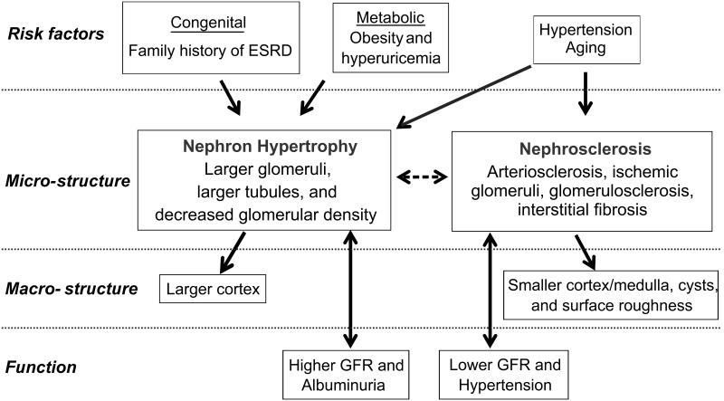
Results: Among 1,520 living kidney donors, nephrosclerosis associated with increased kidney surface roughness, cysts, and smaller cortical to medullary volume ratio. Larger nephron size (nephron hypertrophy) associated with larger cortical volume. Nephron hypertrophy and larger cortical volume associated with higher systolic blood pressure, glomerular filtration rate, and urine albumin excretion; larger body mass index; higher serum uric acid level; and family history of end-stage renal disease. Both nephron hypertrophy and nephrosclerosis associated with older age and mild hypertension. The net effect of both nephron hypertrophy and nephrosclerosis associating with cortical volume was that nephron hypertrophy diminished volume loss with age-related nephrosclerosis and fully negated volume loss with mild hypertension-related nephrosclerosis.
Limitations: Kidney donors are selected on health, restricting the spectrum of pathologic findings. Kidney biopsies in living donors are a small tissue sample leading to imprecise estimates of structural findings.
Conclusions: Among apparently healthy adults, the microstructural findings of nephron hypertrophy and nephrosclerosis differ in their associations with kidney function, macrostructure, and risk factors.
Keywords: Nephrosclerosis; aging; arteriosclerosis; biopsy; chronic kidney disease (CKD) risk factor; contrast computed tomographic (CT) scan; high-resolution imaging; hypertension; kidney function; kidney macrostructure; kidney microstructure; kidney volume; living kidney donor; nephron hypertrophy; subclinical renal pathology.
Copyright © 2016 National Kidney Foundation, Inc. Published by Elsevier Inc. All rights reserved.
Figures
Similar articles
-
Structural and Functional Changes in Human Kidneys with Healthy Aging.J Am Soc Nephrol. 2017 Oct;28(10):2838-2844. doi: 10.1681/ASN.2017040421. Epub 2017 Aug 8. J Am Soc Nephrol. 2017. PMID: 28790143 Free PMC article. Review.
-
Larger nephron size, low nephron number, and nephrosclerosis on biopsy as predictors of kidney function after donating a kidney.Am J Transplant. 2019 Jul;19(7):1989-1998. doi: 10.1111/ajt.15259. Epub 2019 Feb 1. Am J Transplant. 2019. PMID: 30629312 Free PMC article.
-
Nephron hypertrophy and glomerulosclerosis and their association with kidney function and risk factors among living kidney donors.Clin J Am Soc Nephrol. 2014 Nov 7;9(11):1892-902. doi: 10.2215/CJN.02560314. Epub 2014 Oct 15. Clin J Am Soc Nephrol. 2014. PMID: 25318758 Free PMC article.
-
Kidney Structural Features from Living Donors Predict Graft Failure in the Recipient.J Am Soc Nephrol. 2020 Feb;31(2):415-423. doi: 10.1681/ASN.2019090964. Epub 2020 Jan 23. J Am Soc Nephrol. 2020. PMID: 31974271 Free PMC article.
-
Structural and Functional Changes With the Aging Kidney.Adv Chronic Kidney Dis. 2016 Jan;23(1):19-28. doi: 10.1053/j.ackd.2015.08.004. Adv Chronic Kidney Dis. 2016. PMID: 26709059 Free PMC article. Review.
Cited by
-
Structural and Functional Changes in Human Kidneys with Healthy Aging.J Am Soc Nephrol. 2017 Oct;28(10):2838-2844. doi: 10.1681/ASN.2017040421. Epub 2017 Aug 8. J Am Soc Nephrol. 2017. PMID: 28790143 Free PMC article. Review.
-
The Development of a Predictive Model for Postoperative Renal Function in Living Kidney-Transplant Donors.J Clin Med. 2024 Nov 23;13(23):7090. doi: 10.3390/jcm13237090. J Clin Med. 2024. PMID: 39685548 Free PMC article.
-
Changes in Glomerular Volume, Sclerosis, and Ischemia at 5 Years after Kidney Transplantation: Incidence and Correlation with Late Graft Failure.J Am Soc Nephrol. 2023 Feb 1;34(2):346-358. doi: 10.1681/ASN.2022040418. Epub 2022 Nov 17. J Am Soc Nephrol. 2023. PMID: 36396330 Free PMC article.
-
Klotho as a potential predictor of deceased donor kidney transplantation outcomes.Ann Surg Treat Res. 2020 Jun;98(6):332-339. doi: 10.4174/astr.2020.98.6.332. Epub 2020 May 28. Ann Surg Treat Res. 2020. PMID: 32528913 Free PMC article.
-
The conundrums of chronic kidney disease and aging.J Nephrol. 2017 Aug;30(4):477-483. doi: 10.1007/s40620-016-0362-x. Epub 2016 Nov 25. J Nephrol. 2017. PMID: 27885585 Review.
References
-
- Mattix HJ, Hsu CY, Shaykevich S, Curhan G. Use of the albumin/creatinine ratio to detect microalbuminuria: implications of sex and race. J Am Soc Nephrol. 2002;13(4):1034–1039. - PubMed
-
- Hricak H, Cruz C, Romanski R, et al. Renal parenchymal disease: sonographic-histologic correlation. Radiology. 1982;144(1):141–147. - PubMed
-
- Chung EM, Conran RM, Schroeder JW, Rohena-Quinquilla IR, Rooks VJ. From the radiologic pathology archives: pediatric polycystic kidney disease and other ciliopathies: radiologic-pathologic correlation. Radiographics. 2014;34(1):155–178. - PubMed
-
- Rosenfield AT, Siegel NJ. Renal parenchymal disease: histopathologic-sonographic correlation. AJR Am J Roentgenol. 1981;137(4):793–798. - PubMed
Publication types
MeSH terms
Grants and funding
LinkOut - more resources
Full Text Sources
Other Literature Sources
