. 2016 Apr 1;291(14):7465-76.
doi: 10.1074/jbc.M115.700344. Epub 2016 Feb 8.

Structure and Functional Properties of the Active Form of the Proteolytic Complex, ClpP1P2, from Mycobacterium tuberculosis

Affiliations

Structure and Functional Properties of the Active Form of the Proteolytic Complex, ClpP1P2, from Mycobacterium tuberculosis

Mi Li et al. J Biol Chem. .

Abstract

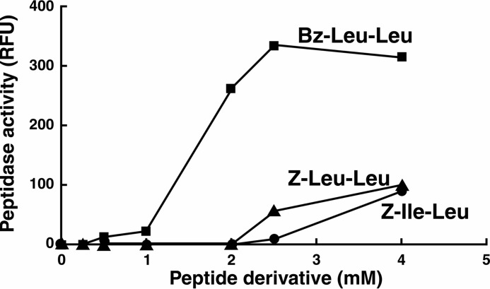
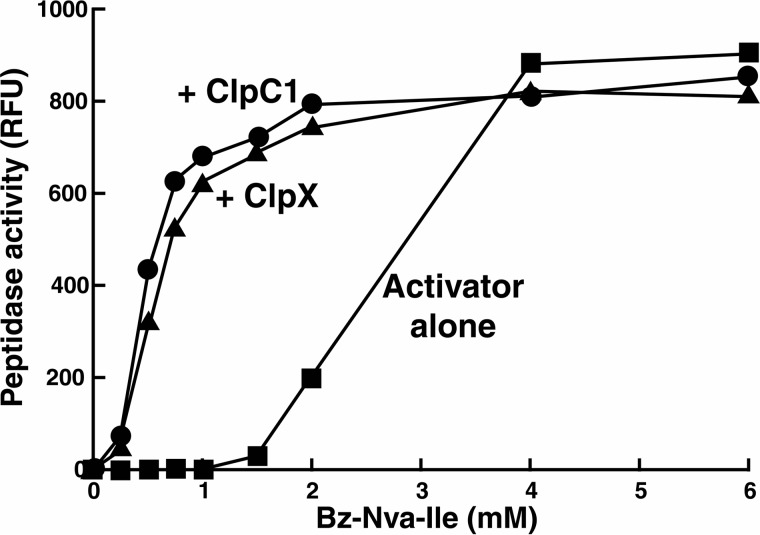
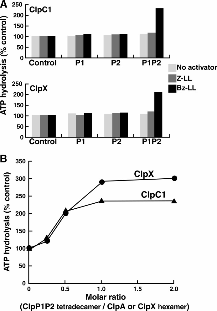
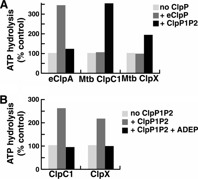
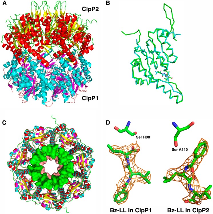
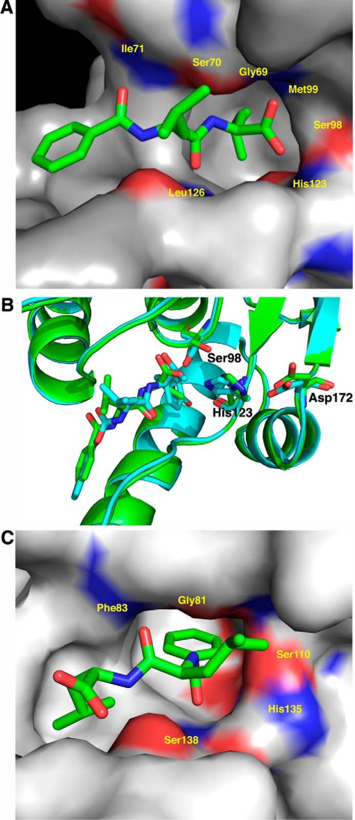
The ClpP protease complex and its regulatory ATPases, ClpC1 and ClpX, inMycobacterium tuberculosis(Mtb) are essential and, therefore, promising drug targets. TheMtbClpP protease consists of two heptameric rings, one composed of ClpP1 and the other of ClpP2 subunits. Formation of the enzymatically active ClpP1P2 complex requires binding of N-blocked dipeptide activators. We have found a new potent activator, benzoyl-leucine-leucine (Bz-LL), that binds with higher affinity and promotes 3-4-fold higher peptidase activity than previous activators. Bz-LL-activated ClpP1P2 specifically stimulates the ATPase activity ofMtbClpC1 and ClpX. The ClpC1P1P2 and ClpXP1P2 complexes exhibit 2-3-fold enhanced ATPase activity, peptide cleavage, and ATP-dependent protein degradation. The crystal structure of ClpP1P2 with bound Bz-LL was determined at a resolution of 3.07 Å and with benzyloxycarbonyl-Leu-Leu (Z-LL) bound at 2.9 Å. Bz-LL was present in all 14 active sites, whereas Z-LL density was not resolved. Surprisingly, Bz-LL adopts opposite orientations in ClpP1 and ClpP2. In ClpP1, Bz-LL binds with the C-terminal leucine side chain in the S1 pocket. One C-terminal oxygen is close to the catalytic serine, whereas the other contacts backbone amides in the oxyanion hole. In ClpP2, Bz-LL binds with the benzoyl group in the S1 pocket, and the peptide hydrogen bonded between parallel β-strands. The ClpP2 axial loops are extended, forming an open axial channel as has been observed with bound ADEP antibiotics. Thus occupancy of the active sites of ClpP allosterically alters sites on the surfaces thereby affecting the association of ClpP1 and ClpP2 rings, interactions with regulatory ATPases, and entry of protein substrates.

Keywords: enzyme mechanism; protease; protein complex; protein degradation; x-ray crystallography.


Figures

FIGURE 1.
FIGURE 1.
Bz-LL is a more potent activator of ClpP1P2 than dipeptides described previously. Hydrolysis of fluorogenic peptide Ac-PKM-amc was assayed at 37 °C as described under “Experimental Procedures.” Dipeptide activators (Bz-Leu-Leu, Z-Leu-Leu, or Z-Ile-Leu) were added at the concentrations shown. The production of fluorescent 7-amino-4-methylcoumarin from Ac-PKM-amc was monitored by the increase in fluorescence at 460 nm (excitation at 380 nm). Each point is the average of 3 measurements, which agreed within 5%. Similar results were obtained in at least three independent experiments. RFU, relative fluorescence.
FIGURE 2.
FIGURE 2.
The presence of ClpC1 or ClpX lowers the concentration of dipeptide activator needed to stimulate peptidase activity of ClpP1P2. Peptidase activity was measured with Ac-PKM-amc as described under “Experimental Procedures” but with varying concentrations of the activator Bz-Nva-Ile. Similar results were obtained with Bz-Leu-Leu (not shown). Where indicated, assay solutions also contained either ClpX or ClpC1, which was added in equimolar concentrations with the ClpP1P2 complex. Each point is the average of three measurements, which agreed within 5%. Similar results were obtained in at least three independent experiments. RFU, relative fluorescence.
FIGURE 3.
FIGURE 3.
Peptide-activated ClpP1P2 stimulates ATPase activities of ClpX and ClpC1. A, ClpP1P2 activates ATP hydrolysis by ClpC1 and ClpX only in the presence of Bz-LL. ATP hydrolysis was measured in a coupled enzymatic assay with pyruvate kinase and lactate dehydrogenase as described under “Experimental Procedures.” To obtain an active ClpP1P2 complex, ClpP1 (5 mg/ml) and ClpP2 (5 mg/ml) were mixed together in the presence of Bz-LL or Z-LL and incubated for 30 min at room temperature. ClpP1P2 was then diluted 50-fold into 100 μl of reaction mixture that contained 10 μg of ClpC1 (top graph) or 5 μg of ClpX (bottom graph) and the dipeptide activators as indicated. ATPase activity was monitored by the decrease in absorbance at 340 nm. Activities obtained when Z-LL or Bz-LL was present are expressed relative to the activity measured in the absence of activator, which was taken as 100% (Control). Specific activities of ClpC1 and ClpX alone were 1.25 and 1.87 μmol/mg/min, respectively. Each point is the average of 3 measurements, which agreed within 5–10%. Similar results were obtained in at least three independent experiments. B, ratio of ClpP1P2 and ClpC1 or ClpX resulting in maximal activation of ATP hydrolysis. ATP hydrolysis by 10 μg of ClpC1 or 5 μg ClpX was measured in 100 μl of reaction buffer as described in panel A. ClpP1P2 was added in varying concentrations, and Bz-LL (2 mm) was present in all assays. ATPase activity in the absence of ClpP1P2 was taken as 100% (control). Each point is the average of 3 measurements, which agreed within 5–10%. Similar results were obtained in at least three independent experiments.
FIGURE 4.
FIGURE 4.
Specificity of interaction between peptide-activated ClpP1P2 and ClpX or ClpC1. A, E. coli or Mtb ClpP protease complexes stimulate ATPases from the same species only. ATPase activity was measured with 5 μg of Mtb ClpC1, Mtb ClpX, or eClpA in 100 μl of reaction mixture as in Fig. 3A. Assay mixtures had either no ClpP, 10 μg of E. coli ClpP, or 10 μg of Mtb ClpP1P2 plus 2 mm Bz-LL. Activity in the absence of ClpP1P2 was taken as 100% (control). Each point is the average of three measurements, which agreed within 5%. Similar results were obtained in at least three independent experiments. B, ADEP blocks the activation of ClpC1 and ClpX ATPases by ClpP1P2. ATPase assays were conducted as in Fig. 3A. Assay mixtures contained 10 μg of ClpC1 or ClpX and either no addition, 10 μg of ClpP1P2 plus 2 mm Bz-LL, or 10 μg of ClpP1P2 plus 2 mm Bz-LL plus 100 μg ADEP2. Activity in the absence of ClpP1P2 was taken as 100% (control). Each point is the average of 3 measurements, which agreed within 5–10%. Similar results were obtained in at least three independent experiments.
FIGURE 5.
FIGURE 5.
Bz-LL stimulates proteolytic activity of ClpP1P2 more than Z-LL in the presence or absence of the regulatory ATPases. A, Bz-LL enhanced the ability of ClpC1 or ClpX to promote protein degradation by ClpP1P2. Degradation of FITC-casein by ClpP1P2 in the presence of ClpC1 and degradation of GFP-SsrA by ClpP1P2 in the presence of ClpX were measured continuously as described under “Experimental Procedures” in buffer A containing either 2 mm Mg-ATP or 100 μm ATPγS and the dipeptide activators as indicated. For ease of comparison, data were normalized to the rate of degradation in the presence of Z-LL, which was taken as 100% (control). Each point is the average of 3 measurements, which agreed within 5–10%. Similar results were obtained in at least three independent experiments. B, Bz-LL promotes degradation of peptides and unfolded proteins by ClpP1P2 in the absence of ATPases. FITC-casein was incubated with a mixture of ClpP1 and ClpP2 in the presence of 2 mm Bz-LL or 5 mm of Z-LL. Degradation was monitored by the decrease in fluorescence of FITC-casein as described under “Experimental Procedures.” Peptidase activity was measured as described in Fig. 1. Protein degradation or peptidase activity in the presence of Z-LL was designated as 100% (control). Each point is the average of 3 measurements, which agreed within 5–10%. Similar results were obtained in at least three independent experiments.
FIGURE 6.
FIGURE 6.
Structural features of Mtb ClpP1P2. A, the tetradecamer is a complex of ClpP1 and ClpP2 heptamers. The mixed tetradecamer formed by joining the heptamers of ClpP1 (cyan helices and magenta β-strands) and ClpP2 (red helices and yellow β-strands) is shown in side view in schematic representation. In ClpP2 the N-terminal β-hairpins are in an extended conformation (yellow and green). B, ribbon representation showing the overlap of ClpP1 and ClpP2 subunits. Backbone atoms between residues 20–185 align with root mean square deviation of 1.2 Å. ClpP2 (green ribbon) has an extended N-terminal β-hairpin that is shorter and disordered in ClpP1 (cyan). ClpP2 also has a bulge in the coiled portion of the alpha-β handle that forms the interface between the heptamers in the tetradecameric complex. The activators are depicted as narrow sticks colored according to the subunits to which they are bound. C, overlap of the ClpP1 and ClpP2 heptamers. Stick representations of ClpP1 and ClpP2 are overlaid. Residues 13–19 of ClpP1 and 15–32 of ClpP2 are shown in surface rendering to illustrate the difference in the axial channel diameter in the two heptamers. D, 2FoFc electron density of the activator bound in ClpP1 and ClpP2. The molecules lie in opposite orientations relative to the catalytic serine shown as sticks.
FIGURE 7.
FIGURE 7.
The activator is bound in opposite orientations in ClpP1 and ClpP2. Subunits are shown in surface rendering and colored red (negative) and blue (positive) to illustrate the electrostatic surfaces in the vicinity of the catalytic serine and histidine residues (shown as sticks). The S1 pocket, which accepts the P1 side chain of bound substrates, is identifiable as a deep gray cavern. A, Bz-LL in ClpP1. Bz-LL (cyan stick) is also held in place by hydrogen bonding with backbone atoms but lies in the opposite orientation with the side chain of leucine in the S1 pocket. One of the C-terminal oxygens of Bz-LL makes weak hydrogen-bonding interactions with the catalytic serine and histidine residues. The other C-terminal oxygen interacts with the oxyanion hole created by the backbone amides from Gly-69 and Met-99. B, Bz-LL in ClpP1 is aligned in the same manner as the covalently bound active site inhibitor, Z-LY-chloromethyl ketone. Overlap of E. coli ClpP with Z-LY-chloromethyl ketone covalently bound at the active site (PDB code 2FZS) (cyan) with Mtb ClpP1 with Bz-LL bound (green). Both ligands make identical backbone contacts with ClpP and engage the catalytic residues. C, Bz-LL in ClpP2. Bz-LL (cyan stick) is held in place by backbone hydrogen bonding and the bonding of the benzoyl moiety in the S1 pocket. The catalytic serine and histidine residues are rotated toward each other, but there are no interactions between Bz-LL and the catalytic residues.

Similar articles

Cited by

References

    1. Porankiewicz J., Wang J., and Clarke A. K. (1999) New insights into the ATP-dependent Clp protease: Escherichia coli and beyond. Mol. Microbiol. 32, 449–458 - PubMed
    1. Katayama-Fujimura Y., Gottesman S., and Maurizi M. R. (1987) A multiple-component, ATP-dependent protease from Escherichia coli. J. Biol. Chem. 262, 4477–4485 - PubMed
    1. Hwang B. J., Park W. J., Chung C. H., and Goldberg A. L. (1987) Escherichia coli contains a soluble ATP-dependent protease (Ti) distinct from protease La. Proc. Natl. Acad. Sci. U.S.A. 84, 5550–5554 - PMC - PubMed
    1. Wang J., Hartling J. A., and Flanagan J. M. (1997) The structure of ClpP at 2.3 Å resolution suggests a model for ATP- dependent proteolysis. Cell 91, 447–456 - PubMed
    1. Kessel M., Maurizi M. R., Kim B., Kocsis E., Trus B. L., Singh S. K., and Steven A. C. (1995) Homology in structural organization between E. coli ClpAP protease and the eukaryotic 26 S proteasome. J. Mol. Biol. 250, 587–594 - PubMed

Publication types

MeSH terms

LinkOut - more resources