. 2016 Feb 15;212(4):389-97.
doi: 10.1083/jcb.201508026. Epub 2016 Feb 8.

Coupling primary and stem cell-derived cardiomyocytes in an in vitro model of cardiac cell therapy

Affiliations

Coupling primary and stem cell-derived cardiomyocytes in an in vitro model of cardiac cell therapy

Yvonne Aratyn-Schaus et al. J Cell Biol. .

Abstract

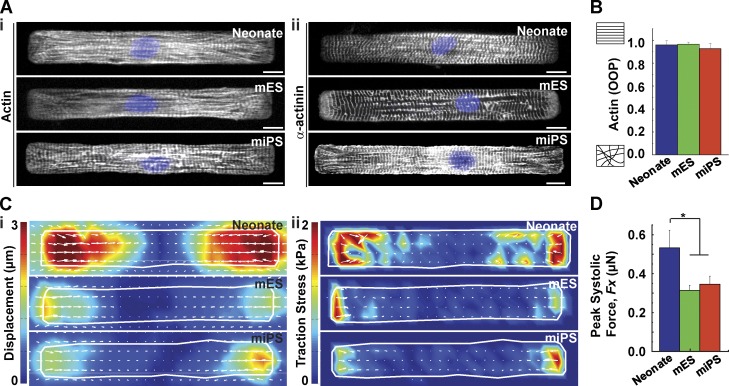
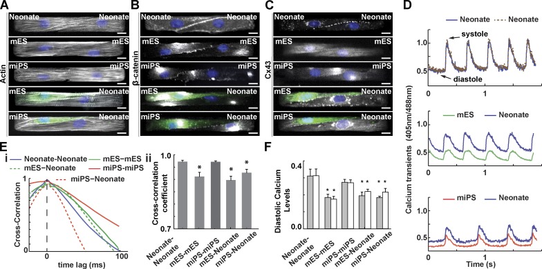
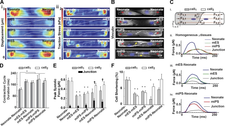
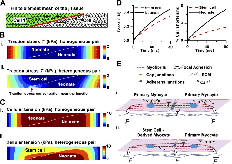
The efficacy of cardiac cell therapy depends on the integration of existing and newly formed cardiomyocytes. Here, we developed a minimal in vitro model of this interface by engineering two cell microtissues (μtissues) containing mouse cardiomyocytes, representing spared myocardium after injury, and cardiomyocytes generated from embryonic and induced pluripotent stem cells, to model newly formed cells. We demonstrated that weaker stem cell-derived myocytes coupled with stronger myocytes to support synchronous contraction, but this arrangement required focal adhesion-like structures near the cell-cell junction that degrade force transmission between cells. Moreover, we developed a computational model of μtissue mechanics to demonstrate that a reduction in isometric tension is sufficient to impair force transmission across the cell-cell boundary. Together, our in vitro and in silico results suggest that mechanotransductive mechanisms may contribute to the modest functional benefits observed in cell-therapy studies by regulating the amount of contractile force effectively transmitted at the junction between newly formed and spared myocytes.


Figures

Figure 1.
Figure 1.
Isolated primary and stem cell–derived cardiac myocytes. (A) Representative images of mononucleated (DAPI, blue) mouse neonate myocytes or mES- or miPS-CMs stained for actin (i) and α-actinin (ii). Bar, 10 µm. (B) Actin OOP as a function of cell type (n = 25, 20, and 9 for neonate, mES, and miPS, respectively). (C) Representative heat maps indicating peak systolic displacement (i) and traction stress (ii). (D) Peak systolic force as a function of cell type (n = 8, 7, and 7 for neonate, mES, and miPS, respectively). Results presented as mean ± SEM. *, P < 0.05.
Figure 2.
Figure 2.
Structural and electrochemical coupling of paired myocytes. Representative images of pairs of mononucleated (DAPI, blue) mouse neonate myocytes or mES or miPS-CMs stained for actin (A), β-catenin (B), and connexin-43 (C). Bar, 10 µm. GFP-tagged (green) mES- or miPS-CMs were used for heterogeneous pairing. (D) Representative ratiometric Ca2+ transients across several contraction cycles for different µtissue types. (E) Normalized cross-correlation functions and cross-correlation coefficients for different µtissue types. (F) Diastolic Ca2+ levels for each cell within a cell pair. Results are presented as mean ± SEM (n = 8, 13, 9, 15, and 6 for neonate-neonate, mES-mES, mES-neonate, miPS-miPS, and miPS-neonate, respectively). *, P < 0.05, statistically significant difference with homogeneous neonate pair.
Figure 3.
Figure 3.
Mechanical coupling of paired myocytes. (A and B) Representative heat maps indicating peak systolic displacement (A, i), traction stress (A, ii), and vinculin-positive (B, gray) adhesions (arrows) for pairs of mononucleated (DAPI, blue) myocytes. Bar, 10 µm. (C) Schematic diagram indicating force calculations for different locations within cell pairs (i). Representative contraction cycles for each cell within homogeneous (ii) and heterogeneous mES-CM (iii) or miPS-CM (iv) pairs. (D) Duration of the contraction cycle as a function of μtissue type. (E) Peak systolic force in different regions within cell pairs. (F) Percentage cell shortening across homogeneous and heterogeneous cell pairs. Results presented as mean ± SEM (n = 7, 12, 8, 7, and 7 for neonate-neonate, mES-mES, mES-neonate, miPS-miPS, and miPS-neonate, respectively). *, P < 0.05, statistically significant differences from the neonate-neonate case for cellular values; , P < 0.05, statistically significant differences from the neonate-neonate case for junctional values.
Figure 4.
Figure 4.
Computational model of μtissue mechanics. (A) Representative finite element mesh of μtissue (cell–cell junction highlighted in red). (B and C) Predicted patterns of local traction (B) and cellular (C) stress at peak systole for homogeneous (i) and heterogeneous (ii) pairs. The red arrow indicates high traction stress beneath the neonate myocyte near the cell–cell junction. (D) Predicted traction force and percentage shortening in heterogeneous cell pairs. (E) Schematic illustration indicates typical tension experienced at substrate adhesions within pairs of neonate cells (i) and heterogeneous (ii) µtissues.

Comment in

References

    1. Abdel-Latif A., Bolli R., Tleyjeh I.M., Montori V.M., Perin E.C., Hornung C.A., Zuba-Surma E.K., Al-Mallah M., and Dawn B.. 2007. Adult bone marrow-derived cells for cardiac repair: A systematic review and meta-analysis. Arch. Intern. Med. 167:989–997. 10.1001/archinte.167.10.989 - DOI - PubMed
    1. Bers D.M. 2001. Excitation-Contraction Coupling and Cardiac Contractile Force. Springer Netherlands, Dordrecht, Netherlands. 237 pp. 10.1007/978-94-010-0658-3 - DOI
    1. Bers D.M. 2002. Cardiac excitation-contraction coupling. Nature. 415:198–205. 10.1038/415198a - DOI - PubMed
    1. Bolli R., Chugh A.R., D’Amario D., Loughran J.H., Stoddard M.F., Ikram S., Beache G.M., Wagner S.G., Leri A., Hosoda T., et al. . 2011. Cardiac stem cells in patients with ischaemic cardiomyopathy (SCIPIO): Initial results of a randomised phase 1 trial. Lancet. 378:1847–1857. 10.1016/S0140-6736(11)61590-0 - DOI - PMC - PubMed
    1. Butler J.P., Tolić-Nørrelykke I.M., Fabry B., and Fredberg J.J.. 2002. Traction fields, moments, and strain energy that cells exert on their surroundings. Am. J. Physiol. Cell Physiol. 282:C595–C605. 10.1152/ajpcell.00270.2001 - DOI - PubMed

Publication types

MeSH terms