. 2016 Feb 23;113(8):2116-21.
doi: 10.1073/pnas.1600101113. Epub 2016 Feb 8.

Structure-function analysis of myomaker domains required for myoblast fusion

Affiliations

Structure-function analysis of myomaker domains required for myoblast fusion

Douglas P Millay et al. Proc Natl Acad Sci U S A. .

Abstract

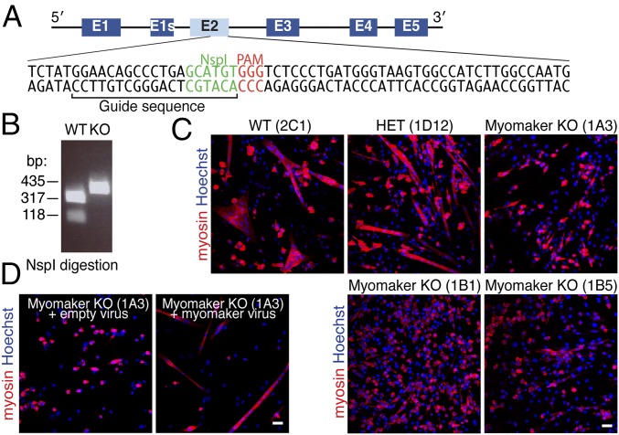
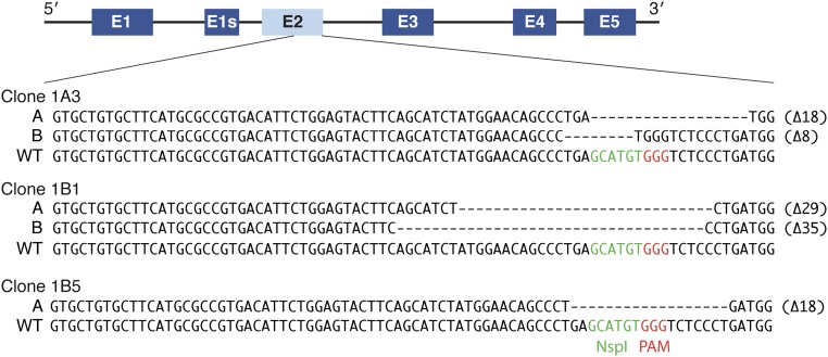
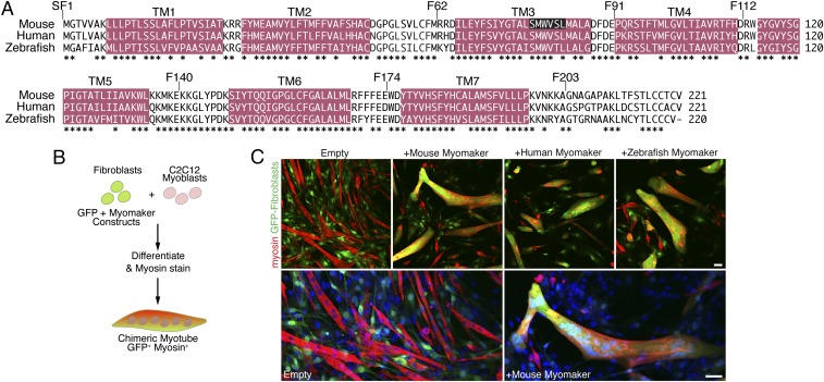
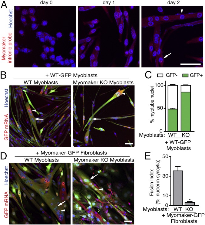
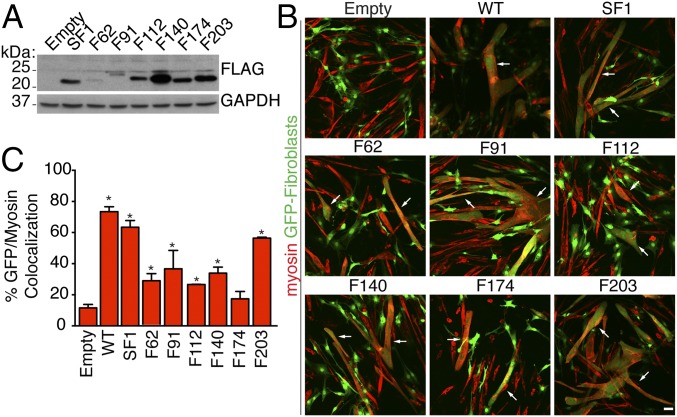
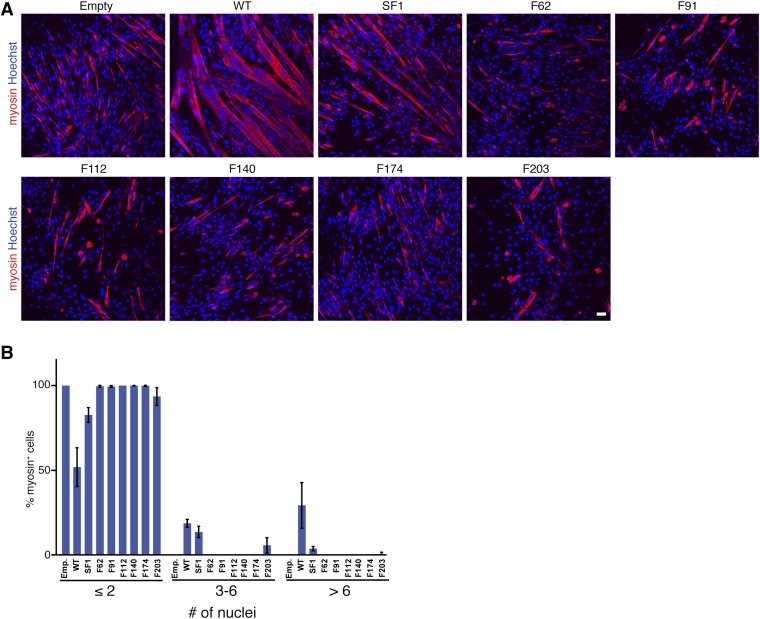
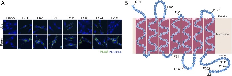
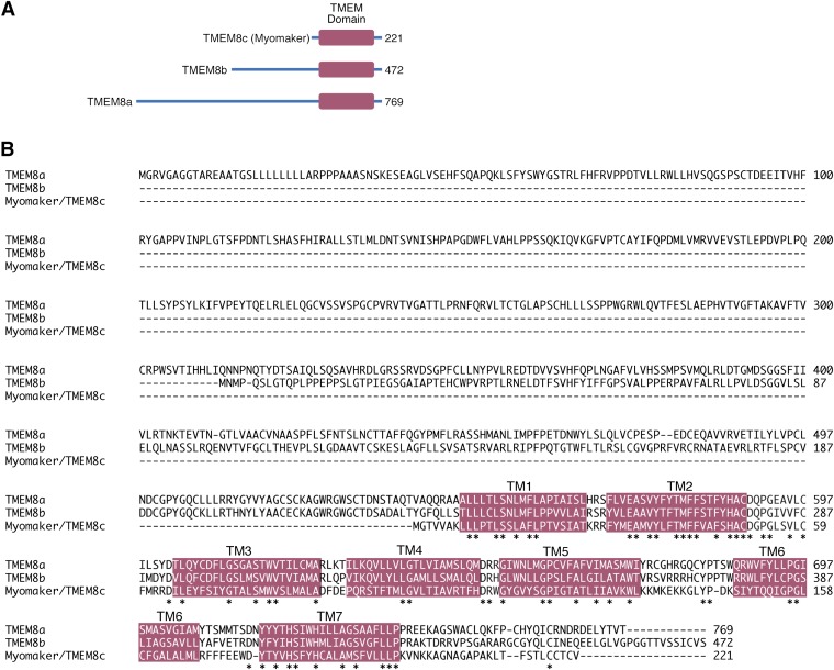
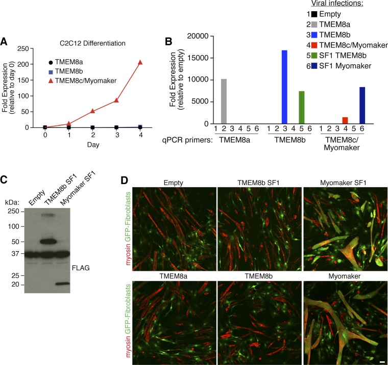
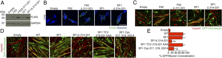
During skeletal muscle development, myoblasts fuse to form multinucleated myofibers. Myomaker [Transmembrane protein 8c (TMEM8c)] is a muscle-specific protein that is essential for myoblast fusion and sufficient to promote fusion of fibroblasts with muscle cells; however, the structure and biochemical properties of this membrane protein have not been explored. Here, we used CRISPR/Cas9 mutagenesis to disrupt myomaker expression in the C2C12 muscle cell line, which resulted in complete blockade to fusion. To define the functional domains of myomaker required to direct fusion, we established a heterologous cell-cell fusion system, in which fibroblasts expressing mutant versions of myomaker were mixed with WT myoblasts. Our data indicate that the majority of myomaker is embedded in the plasma membrane with seven membrane-spanning regions and a required intracellular C-terminal tail. We show that myomaker function is conserved in other mammalian orthologs; however, related family members (TMEM8a and TMEM8b) do not exhibit fusogenic activity. These findings represent an important step toward deciphering the cellular components and mechanisms that control myoblast fusion and muscle formation.

Keywords: CRISPR/Cas9; cell fusion; muscle development; myogenesis.


Conflict of interest statement

The authors declare no conflict of interest.

Figures

Fig. 1.
Fig. 1.
Myomaker disruption in C2C12 cells using CRISPR/Cas9 genome editing. (A) Myomaker locus with exons depicted. Exon 1s is an annotated alternative isoform; however, it is not highly conserved, and we did not investigate it in this study. Exon 2 of myomaker was mutagenized in C2C12 cells after transfection with a vector containing the indicated guide sequence and Cas9. NspI was used to assay for genome disruption at this site. (B) C2C12 clones were genotyped by PCR-amplifying a 435-bp region surrounding the protospacer adjacent motif (PAM) site followed by digestion with NspI. Genomic DNA from myomaker KO clones exhibits an uncut 435-bp fragment because of CRISPR/Cas9-mediated disruption of the NspI site. WT genomic DNA shows 317- and 118-bp bands after NspI digestion. (C) WT (2C1) and HET (1D12) clones formed multinucleated myotubes after 4 d in differentiation media. The three myomaker KO (1A3, 1B1, and 1B5) clones differentiated, shown by myosin staining (red), but failed to fuse. (D) Expression of myomaker using a retrovirus in KO clone 1A3 rescued fusion. Representative images are shown from experiments that were performed twice in duplicate. (Scale bar: 50 μm.)
Fig. S1.
Fig. S1.
Genomic deletion of myomaker exon 2 after CRISPR/Cas 9 mutagenesis. A 435-bp fragment within exon 2 was amplified from genomic DNA from each myomaker KO clonal cell line and ligated into a shuttle vector (TOPO vector). Ten TOPO clones were then sequenced to identify the insertions or deletions after nonhomologous end joining (NHEJ) repair of the double-strand break. The missing nucleotides are indicated by dashes, and the total number of nucleotides disrupted is shown at the end of each line. For clones 1A3 and 1B1, the repair differed for each allele (displayed as A and B sequences). Sequencing of 10 TOPO clones from 1B5 showed the same sequence, suggesting identical repair of both alleles.
Fig. 2.
Fig. 2.
Myomaker orthologs are fusogenic. (A) Amino acid alignment of mouse, human, and zebrafish myomaker proteins. Magenta regions depict significant stretches of hydrophobic amino acids. TM refers to TM domains. Also shown are locations of FLAG epitopes preceding the indicated amino acid (SF1, F62, F91, F112, F140, F174, and F203). Residues deleted in the CRISPR/Cas9 myomaker KO clone 1B5 are displayed with white font and black background. *Sequence conservation. (B) Schematic showing the heterologous cell–cell fusion system. Fibroblasts were infected with GFP and myomaker constructs and mixed with C2C12 myoblasts. The cultures were differentiated for 4 d and then immunostained with myosin antibody as a marker for myocytes. GFP-positive (fibroblast origin), myosin-positive (myoblast origin) chimeric myotubes, in yellow/orange, indicate fusion between the two cell populations. (C) Fibroblasts infected with control (empty) and GFP viruses do not fuse to C2C12 cells. Expression of mouse, human, or zebrafish myomaker in fibroblasts promotes dramatic fusion with muscle. Lower shows enlarged images of cells infected with empty and mouse myomaker retroviruses with Hoechst-stained nuclei. Representative images are shown from experiments that were performed at least three times. (Scale bar: 50 μm.)
Fig. 3.
Fig. 3.
Fusion is most efficient when both cells express myomaker. (A) RNA FISH using a probe specific for myomaker introns was performed on primary WT myoblasts during differentiation. This approach reveals nuclei that are actively transcribing myomaker (red punctae in nuclei). Arrow indicates myoblast nuclei, and arrowhead shows myotube nuclei. (B) WT myoblasts were infected with GFP retrovirus and mixed with either unlabeled WT myoblasts or unlabeled myomaker KO myoblasts. Three days after differentiation, FISH for GFP was performed to track GFP+ nuclei. Arrows depict GFP nuclei, and arrowheads show GFP+ nuclei. (C) Quantitation of the percentage of myotube nuclei that are either GFP+ or GFP reveals that myomaker KO cells fuse less efficiently than WT. (D) Fibroblasts were infected with myomaker and GFP and then mixed with either unlabeled WT myoblasts or unlabeled myomaker KO myoblasts. GFP FISH was performed 3 d after differentiation. Arrows depict GFP nuclei, and arrowheads show GFP+ nuclei. (E) Quantitation of fusion index reveals that WT myoblasts fuse to fibroblasts more efficiently than KO myoblasts. Representative images are shown from experiments that were performed at least three times. (Scale bar: 50 μm.) *P < 0.05 compared with the WT.
Fig. 4.
Fig. 4.
Myomaker epitope-tagged constructs are expressed and functional. (A) Protein extracts from fibroblasts expressing each FLAG-tagged version of myomaker (depicted in Fig. 2A) were analyzed by immunoblotting using an FLAG antibody. Empty-infected fibroblasts were used as a control. Each myomaker-FLAG construct was expressed at varying levels. GAPDH was a loading control. (B) Fibroblasts coinfected with myomaker-FLAG constructs and GFP can fuse with C2C12 cells. Arrows indicate GFP+ myosin+ structures. (C) Quantitation of heterologous fusion through analyzing the colocalization of GFP (fibroblasts) and myosin (myocytes) using CellProfiler. Each tagged version of myomaker exhibits different levels of expression and function. Representative images are shown from experiments that were performed at least three times. (Scale bar: 50 μm.) *P < 0.05 compared with empty.
Fig. S2.
Fig. S2.
Myomaker epitope-tagged constructs do not rescue fusion defect in myomaker KO myoblasts. (A) Myomaker KO myoblasts were infected with various myomaker-FLAG constructs and induced to differentiate. The cultures were stained with myosin antibody and Hoechst to assay fusion. (B) Quantitation of fusion induced by each construct shows a lack of fusion induction by the FLAG constructs. SF1 and F203 do exhibit fusion ability, although they decreased compared with WT. Representative images are shown from experiments that were performed at least three times. Emp., empty. (Scale bar: 50 μm.)
Fig. 5.
Fig. 5.
Determination of myomaker membrane topology. (A) C2C12 cells were infected with myomaker-FLAG retroviral constructs and allowed to differentiate for 1 d. One set of cells was immunostained on ice with a FLAG antibody before fixation or permeabilization (Live), whereas a separate culture was stained after fixation and permeabilization (Perm.). SF1, F62, F112, and F174 exhibited surface staining in live cells, indicating the presence of the epitope on the cell surface. Each construct displayed intracellular punctate localization in permeabilized cells. Representative images are shown from experiments that were performed at least three times. (Scale bar: 50 μm.) (B) Model of myomaker topology based on the positive epitopes in live staining. According to this model, myomaker contains seven TM domains with a 25-aa intracellular C-terminal domain.
Fig. S3.
Fig. S3.
TMEM8a, TMEM8b, and TMEM8c/myomaker sequence comparison. (A) TMEM8a and TMEM8b contain longer N-terminal regions than TMEM8c/myomaker. The number of amino acid residues for each protein is shown. (B) Amino acid alignment between the three TMEM8 family members. Magenta regions depict significant stretches of hydrophobic amino acids. TM refers to TM domains. The majority of homology is localized to the TM domains between the three proteins. *Sequence conservation.
Fig. S4.
Fig. S4.
TMEM8a and TMEM8b differ functionally from myomaker. (A) Quantitative RT-PCR analysis for TMEM8a, TMEM8b, and TMEM8c/myomaker transcripts in C2C12 cells after the indicated day of differentiation. Expression levels were normalized to 18S and expressed as fold change relative to day 0. (B) Quantitative PCR (qPCR) analysis for TMEM8a, TMEM8b, and TMEM8c/myomaker in fibroblasts after infection with empty, TMEM8a, TMEM8b, TMEM8c/myomaker, SF1 TMEM8b, and SF1 myomaker virus. Expression levels were normalized to 18S and expressed as fold change relative to empty-infected cells. (C) FLAG Western blot analysis shows expression of epitope-tagged versions of TMEM8b and myomaker in fibroblasts. (D) The heterologous cell fusion system reveals that fibroblasts infected with empty, TMEM8a, TMEM8b, or TMEM8b SF1 virus do not fuse to C2C12 cells. Fibroblasts containing myomaker or myomaker SF1 readily fuse. Representative images are shown from experiments that were performed at least three times.
Fig. 6.
Fig. 6.
The intracellular C-terminal region of myomaker is required for fusogenic activity. (A) Viruses expressing two independent C-terminal deletion myomaker-FLAG mutants were generated. The final 11 amino acids of F62 (F62Δ211–221) and the last 8 amino acids of SF1 (SF1Δ214–221) were deleted. Expression of each virus was assessed by FLAG immunoblotting. GAPDH was used as a loading control. (B) Myomaker proteins that contained a shorter C terminus were expressed on the membrane as determined by staining live cells after viral transduction. (C) Fibroblasts that express F62Δ211–221 and SF1Δ214–221 do not fuse to myoblasts, whereas F62 and SF1 exhibit robust fusogenic activity. (D) Constructs containing mutations in the final TCV, a PDZ-binding motif, of myomaker (SF1 TCV 219–221 AAA) and the C-terminal cysteines (SF1 Cys-217, 218, and 220 A) were tested for function in the heterologous fusion system. (E) Quantitation of the fusion between fibroblasts and muscle cells. Representative images are shown from experiments that were performed at least three times. (Scale bar: 50 μm.) *P < 0.05 compared with empty; #P < 0.05 compared with SF1.

References

    1. Chen EH, Olson EN. Unveiling the mechanisms of cell-cell fusion. Science. 2005;308(5720):369–373. - PubMed
    1. Abmayr SM, Pavlath GK. Myoblast fusion: Lessons from flies and mice. Development. 2012;139(4):641–656. - PMC - PubMed
    1. Sens KL, et al. An invasive podosome-like structure promotes fusion pore formation during myoblast fusion. J Cell Biol. 2010;191(5):1013–1027. - PMC - PubMed
    1. Dhanyasi N, et al. Surface apposition and multiple cell contacts promote myoblast fusion in Drosophila flight muscles. J Cell Biol. 2015;211(1):191–203. - PMC - PubMed
    1. Laurin M, et al. The atypical Rac activator Dock180 (Dock1) regulates myoblast fusion in vivo. Proc Natl Acad Sci USA. 2008;105(40):15446–15451. - PMC - PubMed

Publication types

MeSH terms