. 2016 Feb 23;113(8):E1064-73.
doi: 10.1073/pnas.1519730113. Epub 2016 Feb 8.

Positive-strand RNA viruses stimulate host phosphatidylcholine synthesis at viral replication sites

Affiliations

Positive-strand RNA viruses stimulate host phosphatidylcholine synthesis at viral replication sites

Jiantao Zhang et al. Proc Natl Acad Sci U S A. .

Abstract

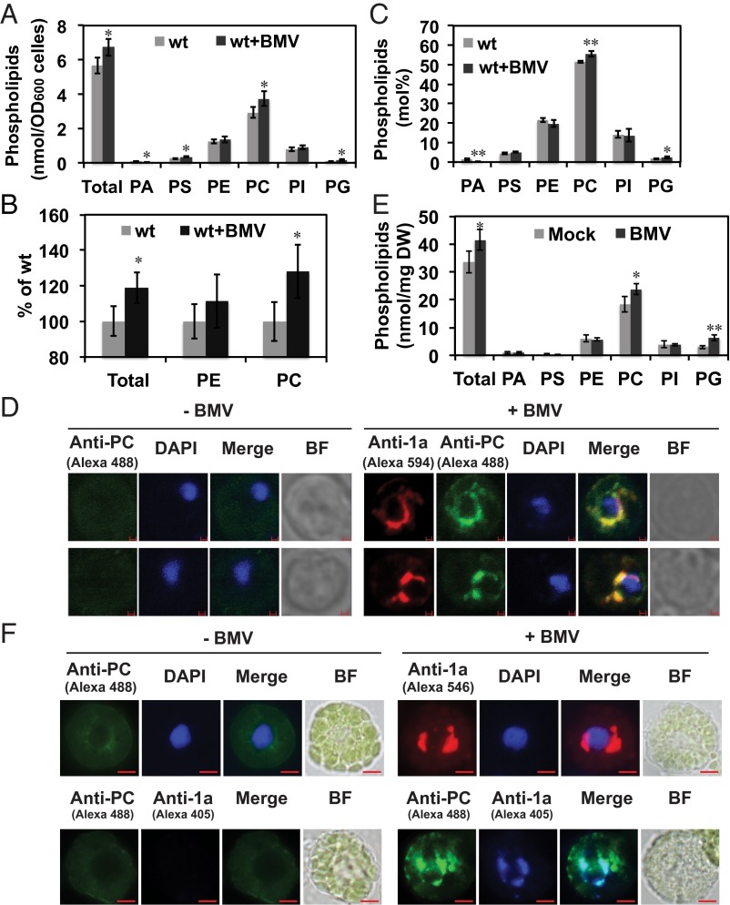
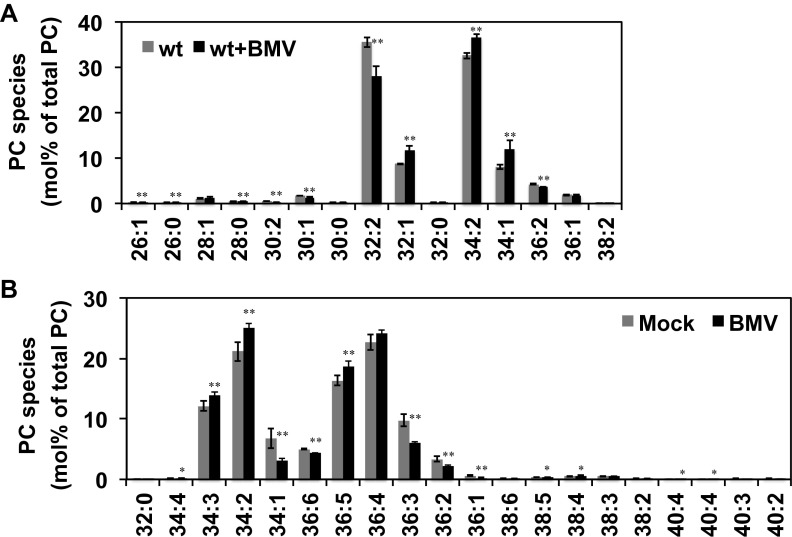
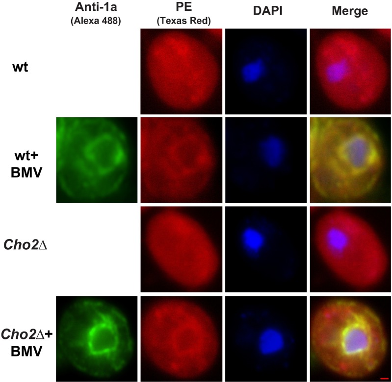
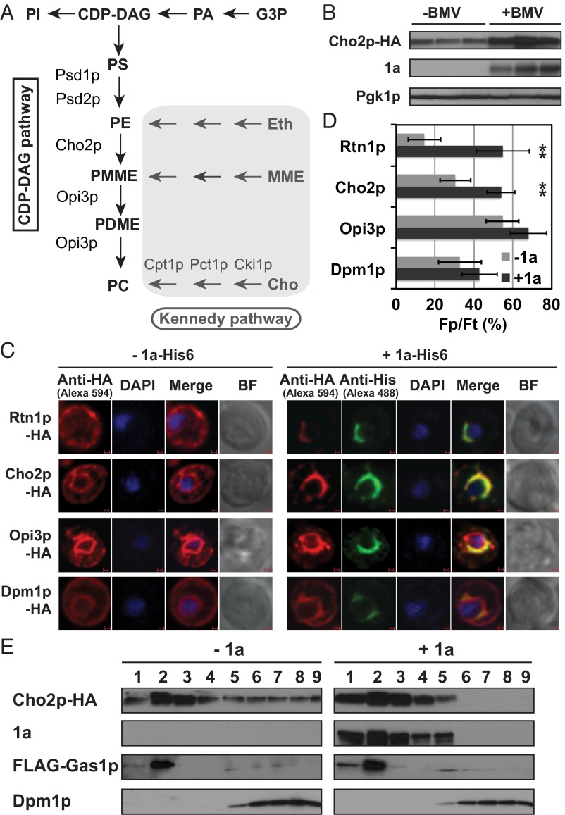
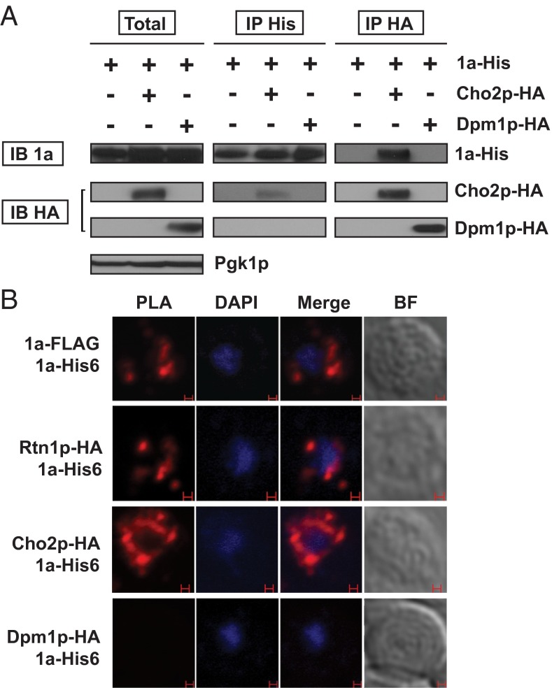
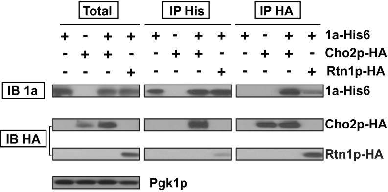
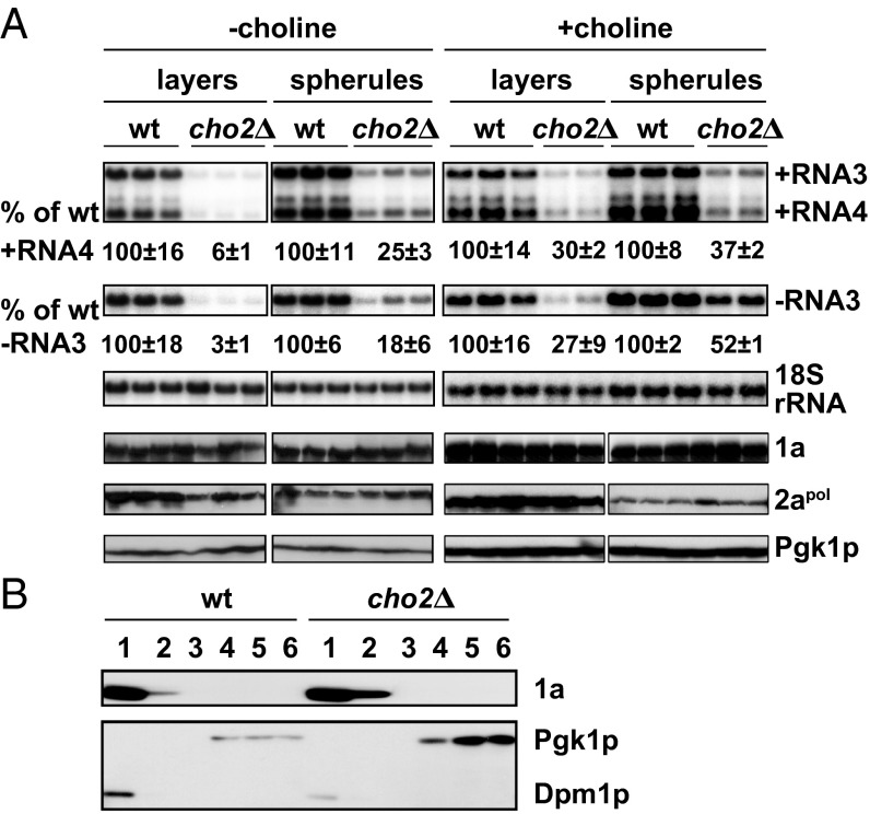
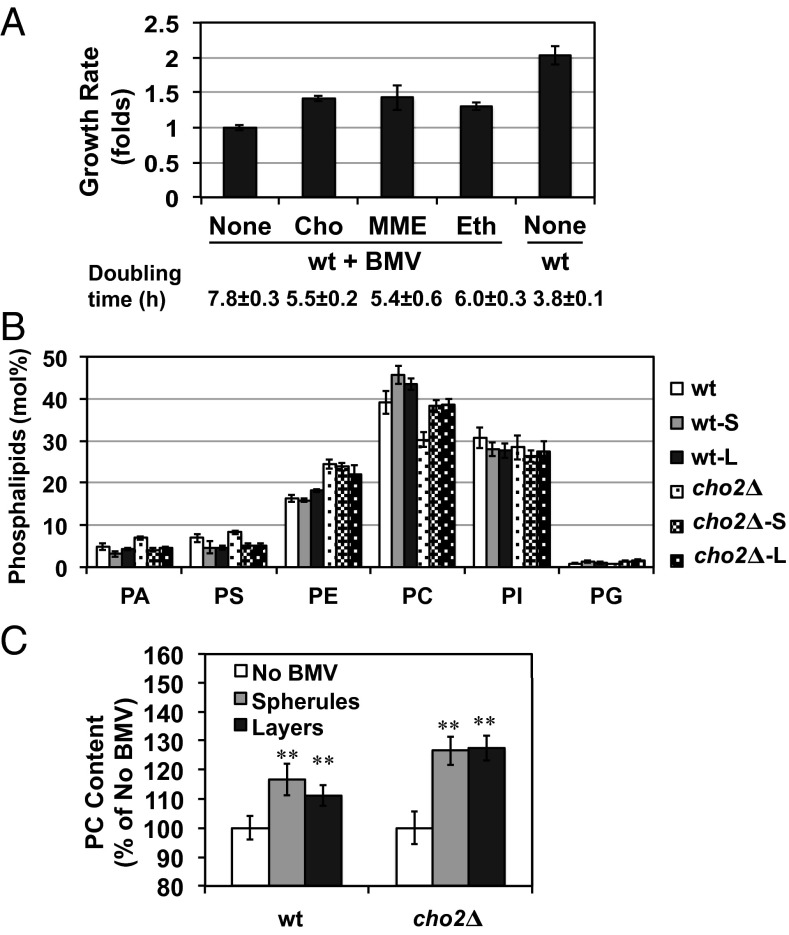
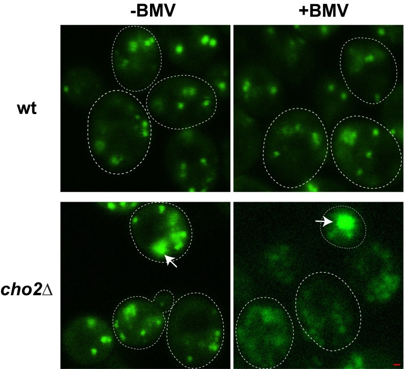
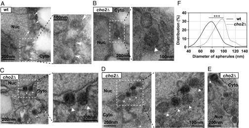
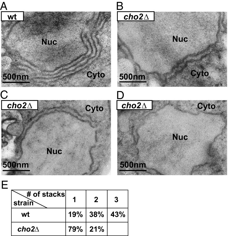
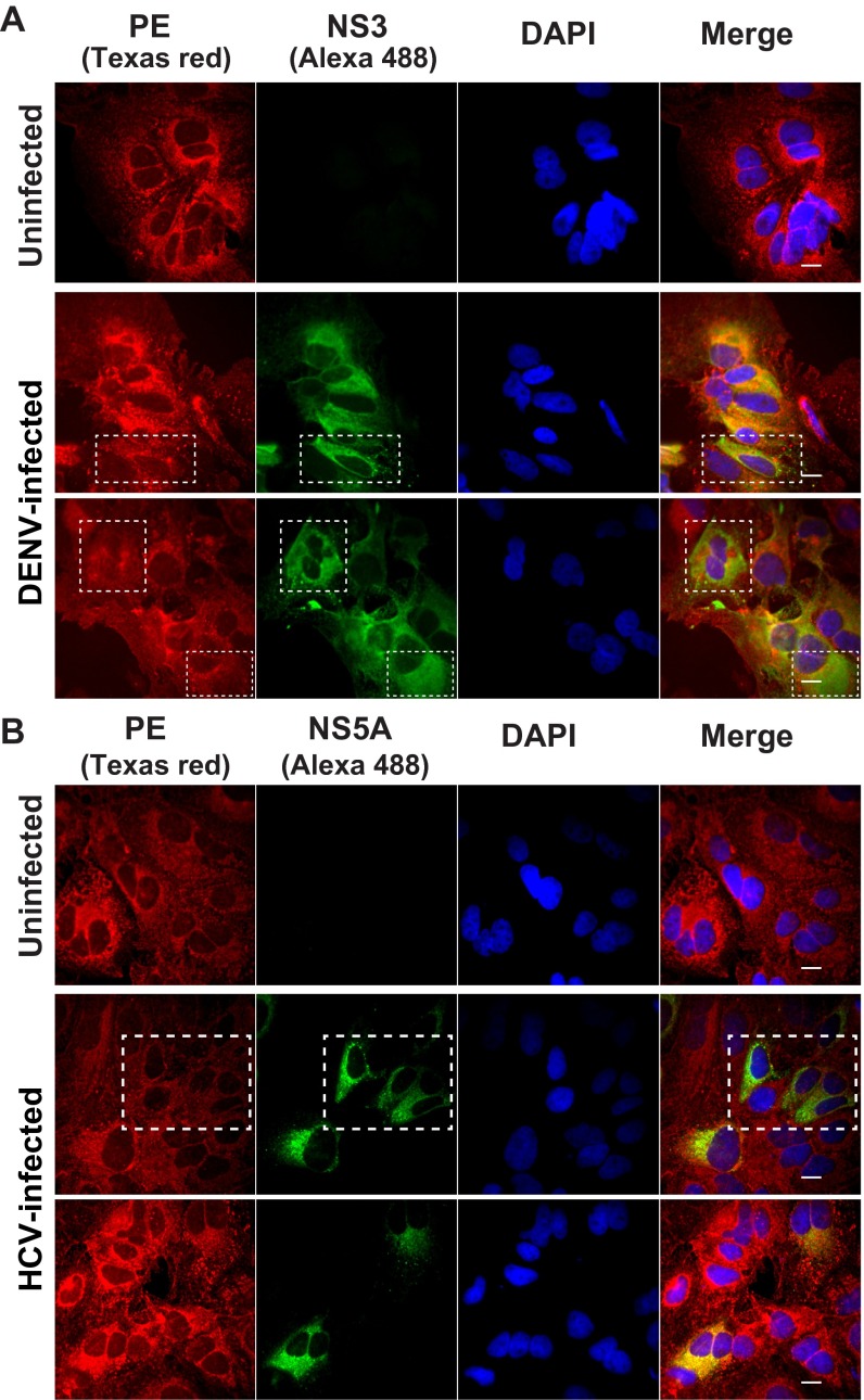
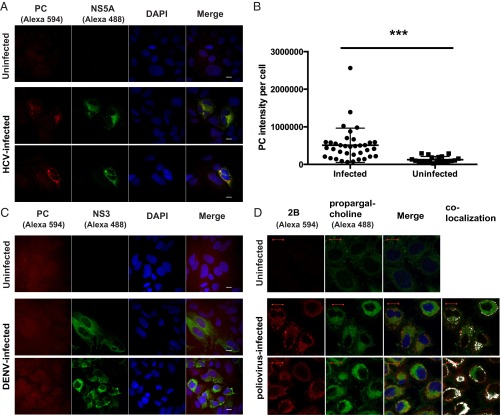
All positive-strand RNA viruses reorganize host intracellular membranes to assemble their viral replication complexes (VRCs); however, how these viruses modulate host lipid metabolism to accommodate such membrane proliferation and rearrangements is not well defined. We show that a significantly increased phosphatidylcholine (PC) content is associated with brome mosaic virus (BMV) replication in both natural host barley and alternate host yeast based on a lipidomic analysis. Enhanced PC levels are primarily associated with the perinuclear ER membrane, where BMV replication takes place. More specifically, BMV replication protein 1a interacts with and recruits Cho2p (choline requiring 2), a host enzyme involved in PC synthesis, to the site of viral replication. These results suggest that PC synthesized at the site of VRC assembly, not the transport of existing PC, is responsible for the enhanced accumulation. Blocking PC synthesis by deleting the CHO2 gene resulted in VRCs with wider diameters than those in wild-type cells; however, BMV replication was significantly inhibited, highlighting the critical role of PC in VRC formation and viral replication. We further show that enhanced PC levels also accumulate at the replication sites of hepatitis C virus and poliovirus, revealing a conserved feature among a group of positive-strand RNA viruses. Our work also highlights a potential broad-spectrum antiviral strategy that would disrupt PC synthesis at the sites of viral replication but would not alter cellular processes.

Keywords: phospholipids; positive-strand RNA viruses; viral replication complexes; virus control; virus–host interactions.


Conflict of interest statement

The authors declare no conflict of interest.

Figures

Fig. 1.
Fig. 1.
BMV promotes PC accumulation at the sites of viral replication. (A) Molar amount of phospholipids in yeast cells in the absence (WT) or presence of BMV replication (WT+BMV). (B) Percentage change in total phospholipids, PE, and PC content based on the molar amount. (C) The relative level of each phospholipid class (molarity percentage, mol%) without or with BMV replication. *P < 0.05; **P < 0.01. (D) IF confocal microscopic images of PC distribution. PC was detected by using an anti-PC mAb (JE-1) and a secondary antibody conjugated to Alexa Fluor 488. BMV 1a was detected with an anti-1a antiserum and a secondary antibody conjugated to Alexa Fluor 594. The yellow color in the merged images indicates the colocalization of PC and 1a signals. The nucleus was stained with DAPI. (Scale bar, 1 μm.) (E) Molar amount of phospholipids in mock- or BMV-inoculated barley plants. (F) PC distribution in barley protoplasts prepared from mock- and BMV-infected systemic leaves. 1a was detected using an anti-1a antiserum. The secondary antibodies for detecting 1a were conjugated to Alexa Fluor 546 or 405 in the Top or Bottom panel, respectively. (Scale bar, 100 μm.)
Fig. S1.
Fig. S1.
The altered host PC accumulation during BMV replication. Shown are profiles of PC species in the absence (WT or mock) and presence of BMV replication (WT+BMV or BMV) in yeast (A) and in barley plants (B). *P < 0.05; **P < 0.01.
Fig. S2.
Fig. S2.
Distribution of PE signals. Shown is the distribution of PE in WT or cho2Δ mutant in the absence (WT or cho2Δ) or presence of BMV replication (WT+BMV or cho2Δ+BMV). PE was detected by biotin-conjugated duromycin peptide and followed by Texas Red-conjugated streptavidin. 1a was detected as in Fig. 1. The nucleus was stained with DAPI. (Scale bar, 5 μm.)
Fig. 2.
Fig. 2.
BMV redistributes Cho2p to the viral replication sites. (A) Diagram of the CDP–DAG and Kennedy pathways involved in PC synthesis. Key enzymes are listed. (B) Accumulated Cho2p, 1a, and pgk1p in the absence or presence of BMV replication. Cho2p was tagged with HA and expressed from its endogenous promoter in cho2Δ cells, and Pgk1p served as a loading control. (C) IF images showing the distributions of host Rtn1p, Cho2p, Opi3p, and Dpm1p in the absence or presence of 1a. (Scale bar, 1 μm.) (D) Percentage of the fluorescent signals [Fp/Ft (%)] of each host protein localized to the perinuclear ER (Fp) versus total fluorescence in the cell (Ft) in the absence or presence of 1a. **P < 0.01. (E) Cho2p is enriched in the DRM fractions in the presence of 1a. Gas1p and Dpm1p serve as positive and negative controls, respectively.
Fig. 3.
Fig. 3.
BMV 1a interacts with Cho2p. (A) Co-IP of 1a-His6 and Cho2p-HA. Cell lysates were subjected to IP using anti-His mAb (IP His) or anti-HA pAb (IP HA), followed by Western blotting using anti-HA (IB HA) or anti-His (IB His) antibodies, respectively. Dpm1p served as a negative control. (B) In situ detection of the 1a–Cho2p interaction via PLA. In cells expressing various protein pairs, PLA signals (red dots) were detected by incubating fixed yeast spheroplasts with anti-His mAb and anti-FLAG pAb or anti-HA pAb following the protocol detailed in Experimental Procedures. The interactions between 1a-FLAG and 1a-His6 as well as Rtn1p-HA and 1a-His6 served as positive controls. The Dpm1p-HA and 1a-His6 pair served as a negative control. (Scale bar, 1 μm.)
Fig. S3.
Fig. S3.
Co-IP of 1a-His6 with Cho2p-HA or Rtn1p-HA. The cell lysate with Cho2p-HA or Rtn1p-HA alone or along with 1a-His6 was subjected to immunoprecipitation using anti-His mAb (IP His) or anti-HA pAb (IP HA), followed by Western blotting using anti-HA (IB HA) or anti-His (IB His) antibodies, respectively. Rtn1p-HA served as a positive control.
Fig. 4.
Fig. 4.
BMV replication is blocked in cho2Δ cells. (A) BMV RNA replication was measured in WT or cho2Δ cells under conditions that led to the formation of double-membrane layers or spherules in the absence or presence of choline. Positive- and negative-strand viral RNAs were detected using BMV strand-specific probes. 18S rRNA served as a loading control. Total proteins were extracted from the same numbers of cells and subjected to Western blotting with anti-1a or -2apol antibodies. Pgk1p served as a loading control. (B) 1a associated similarly with membranes in both WT and cho2Δ cells. 1a was detected in the membrane fractions along with positive control Dpm1p after an iodixanol density gradient. Pgk1p served as a control for soluble proteins.
Fig. 5.
Fig. 5.
BMV induces PC synthesis via the Kennedy pathway. (A) Growth rate of WT cells in the media supplemented with choline, MME, or Eth in the presence of BMV replication. (B) Phospholipid compositions (mol%) in WT or cho2Δ cells with supplemented choline in the absence and presence of BMV replication. S and L, BMV replication as spherule- or layer-replication complexes. (C) Relative PC levels (mol%) in WT and cho2Δ cells without or with BMV replication in the presence of choline to activate the Kennedy pathway; **P < 0.01.
Fig. S4.
Fig. S4.
Illustration of expression cassettes for BMV 1a, 2apol, and RNA3 in yeast. (A) BMV 1a is under the control of a strong GAL1 promoter in both pB1YT3 and pB12VG1. 2apol is expressed under a weak GAL10 promoter in pB12VG1 or a strong GAL1 promoter in pB2YT5, resulting in the formation of spherule or layer replication complexes, respectively, along with BMV 1a. (B) BMV RNA3 is transcribed under a Cupper promoter. However, no copper was supplemented to maintain the minimum transcription of RNA3. A 4-bp insertion in CP coding sequence abolishes CP synthesis, and thus, no viral particles will be produced. (C) Diagram of BMV replication process in yeast. Along with host factors, BMV 1a and 2apol produce (–)RNA3 and (+)RNA4 and amplify (+)RNA3 by using (+)RNA3 transcript as a template.
Fig. S5.
Fig. S5.
Distribution of lipid droplets in WT and cho2Δ cells. WT or cho2Δ cells in the absence or presence of BMV replication were grown to early stationary stage and stained with Nile red for lipid droplets (LDs). Arrows point to supersized LDs in cho2Δ cells. The nucleus was stained by DAPI. (Scale bar, 5 μm.)
Fig. 6.
Fig. 6.
Enlarged spherules are formed in cho2Δ cells. Electron microscopic images of WT (A) or cho2Δ cells (B–E) expressing BMV 1a are shown. Arrows in A point to spherules, and arrowheads in B–E point out spherules with a wider diameter. Images on the Right are higher magnification micrographs of the boxed areas from the images on the Left. (F) Distribution of spherule diameters in WT and cho2Δ cells. ***P < 0.001. Cyto, cytoplasm; Nuc, nucleus.
Fig. 7.
Fig. 7.
Decreased stacks of double-membrane layers are assembled in cho2Δ cells. Electron micrographs of WT (A) or cho2Δ (B–D) cells expressing BMV 1a and high levels of 2apol are shown. (E) Percentage of cells with visible layer replication complexes that contained a different number of stacked membranes. Cyto, cytoplasm; Nuc, nucleus.
Fig. S6.
Fig. S6.
Distribution of PE during DENV or HCV infection. PE was detected by biotin-conjugated duromycin peptide and followed by Texas Red-conjugated streptavidin in the absence and presence of DENV (A) or HCV (B). The boxed cells indicated high levels of signals of viral proteins but not that of PE. The nucleus was stained by DAPI. (Scale bar, 13.5 μm.)
Fig. 8.
Fig. 8.
Newly synthesized PC colocalizes with viral proteins associated with the VRCs of HCV and poliovirus. (A) Huh-7.5 cells were infected with HCV (JC1) at an MOI of 0.05 for 48 h, fixed with 4% (vol/vol) paraformaldehyde, and permeabilized with 0.1% TX100. Cells were then incubated with anti-HCV NS5A mAb (9E10, IgG) and followed by anti-mouse IgG conjugated with Alexa Fluor 488. Cells were then reblocked, incubated with JE1, and followed by anti-mouse IgM (u chain) conjugated to Alexa Fluor 594. The yellow color in the merged images indicates the colocalization of PC and NS5A. DAPI stains the nucleus. (Scale bar, 13.5 μm.) (B) Comparison of PC signal intensity per cell in mock- and HCV-infected Huh-7.5 cells. ***P < 0.001. (C) Huh-7.5 cells were infected with DENV-2 at an MOI of 1 for 48 h. Cells were processed as those of infected by HCV. PC was detected using JE-1, and DENV NS3 was detected by an anti-NS3 antiserum. (Scale bar, 13.5 μm.) (D) HeLa cells were infected with poliovirus type I Mahoney at an MOI of 10. Growth medium with 200 μM of propargal-choline was added at 4 h postinfection and incubated for 1 h. Propargal-choline–labeled PCs were detected by Alexa Fluor 594-conjugated azide. Poliovirus 2B was detected by an anti-2B mAb, and the nucleus was stained with Hoechst-33342. The colocalization panel shows the colocalized red and green pixels of 2B and PC identified with ImageJ software. (Scale bar, 10 μm.)

Similar articles

Cited by

References

    1. Diaz A, Wang X. Bromovirus-induced remodeling of host membranes during viral RNA replication. Curr Opin Virol. 2014;9(12):104–110. - PubMed
    1. Wang X, Ahlquist P. Brome mosaic virus (Bromoviridae) In: Mahy BWJ, van Regenmortel MHV, editors. Encyclopedia of Virology. 3rd Ed. Elsevier; Oxford, UK: 2008. pp. 381–386.
    1. Ahola T, Ahlquist P. Putative RNA capping activities encoded by brome mosaic virus: methylation and covalent binding of guanylate by replicase protein 1a. J Virol. 1999;73(12):10061–10069. - PMC - PubMed
    1. Kong F, Sivakumaran K, Kao C. The N-terminal half of the brome mosaic virus 1a protein has RNA capping-associated activities: Specificity for GTP and S-adenosylmethionine. Virology. 1999;259(1):200–210. - PubMed
    1. Wang X, et al. Brome mosaic virus 1a nucleoside triphosphatase/helicase domain plays crucial roles in recruiting RNA replication templates. J Virol. 2005;79(21):13747–13758. - PMC - PubMed

Publication types

MeSH terms

Substances

LinkOut - more resources