. 2016 Feb 23;113(8):2306-11.
doi: 10.1073/pnas.1516732113. Epub 2016 Feb 8.

Titin strain contributes to the Frank-Starling law of the heart by structural rearrangements of both thin- and thick-filament proteins

Affiliations

Titin strain contributes to the Frank-Starling law of the heart by structural rearrangements of both thin- and thick-filament proteins

Younss Ait-Mou et al. Proc Natl Acad Sci U S A. .

Abstract

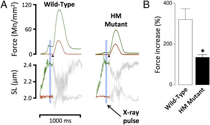
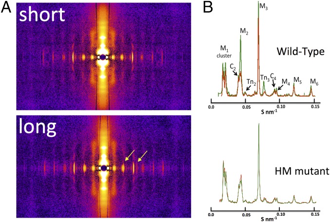
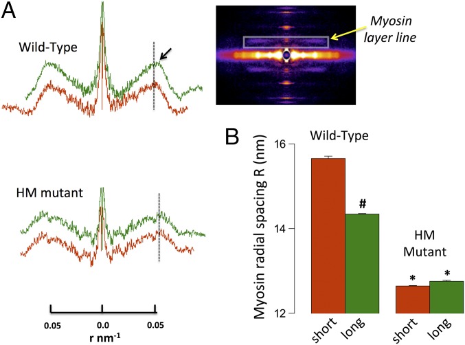
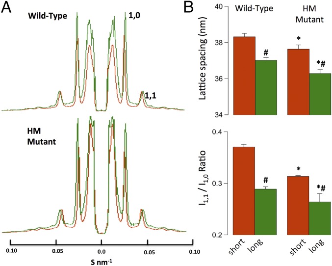
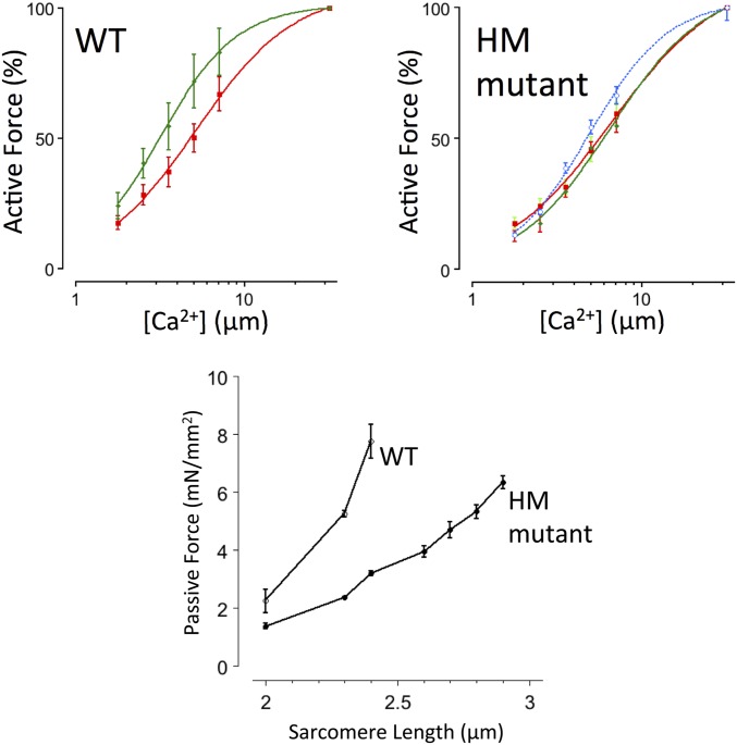
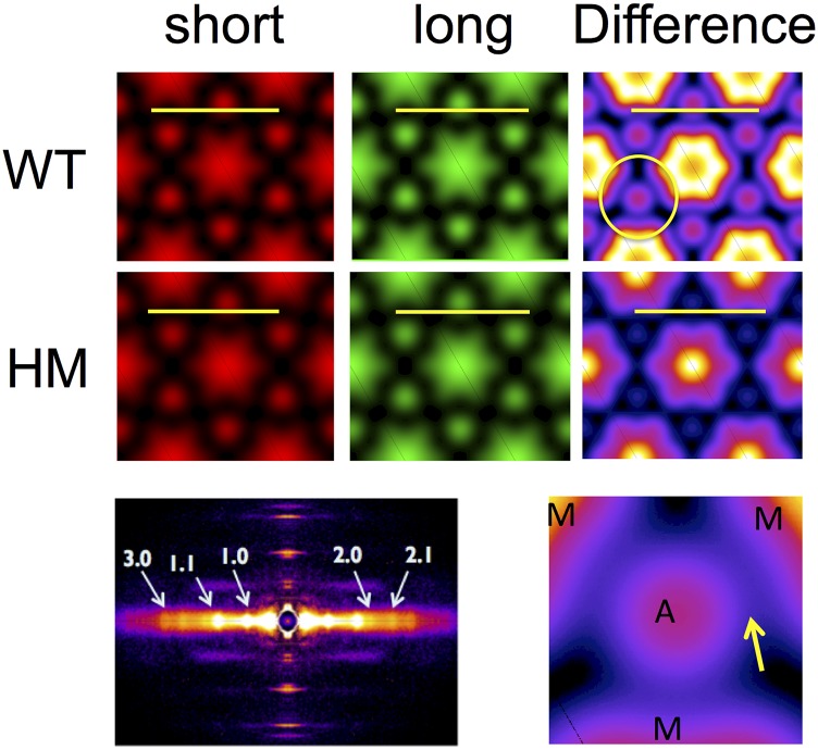
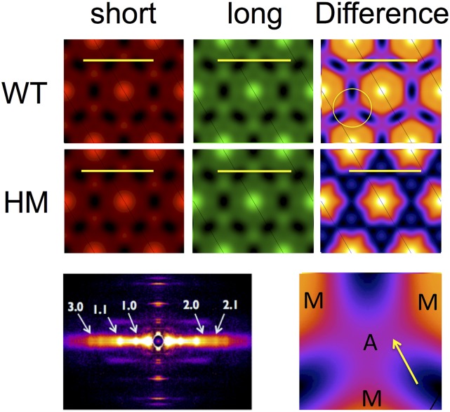
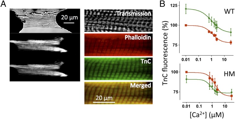
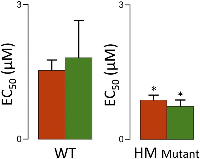
The Frank-Starling mechanism of the heart is due, in part, to modulation of myofilament Ca(2+) sensitivity by sarcomere length (SL) [length-dependent activation (LDA)]. The molecular mechanism(s) that underlie LDA are unknown. Recent evidence has implicated the giant protein titin in this cellular process, possibly by positioning the myosin head closer to actin. To clarify the role of titin strain in LDA, we isolated myocardium from either WT or homozygous mutant (HM) rats that express a giant splice isoform of titin, and subjected the muscles to stretch from 2.0 to 2.4 μm of SL. Upon stretch, HM compared with WT muscles displayed reduced passive force, twitch force, and myofilament LDA. Time-resolved small-angle X-ray diffraction measurements of WT twitching muscles during diastole revealed stretch-induced increases in the intensity of myosin (M2 and M6) and troponin (Tn3) reflections, as well as a reduction in cross-bridge radial spacing. Independent fluorescent probe analyses in relaxed permeabilized myocytes corroborated these findings. X-ray electron density reconstruction revealed increased mass/ordering in both thick and thin filaments. The SL-dependent changes in structure observed in WT myocardium were absent in HM myocardium. Overall, our results reveal a correlation between titin strain and the Frank-Starling mechanism. The molecular basis underlying this phenomenon appears not to involve interfilament spacing or movement of myosin toward actin but, rather, sarcomere stretch-induced simultaneous structural rearrangements within both thin and thick filaments that correlate with titin strain and myofilament LDA.

Keywords: fluorescent probes; myofilament length-dependent activation; passive force; rat; small-angle X-ray diffraction.


Conflict of interest statement

The authors declare no conflict of interest.

Figures

Fig. 1.
Fig. 1.
Impact of titin length on cardiac muscle function. Cardiac muscles were isolated from WT or HM rats and electrically stimulated (arrowheads). (A) Force and SL recordings; SL in the diastolic phase was either maintained at SL = 2.0 μm (red) or increased transiently to SL = 2.4 μm (green). (B) Average percentage increase of twitch force upon stretch (*P < 0.05 WT vs. HM).
Fig. 2.
Fig. 2.
Two-dimensional X-ray diffraction and meridional analysis. (A) Representative 2D X-ray diffraction patterns in WT at short and long SL. Stretch-induced distinct alterations in the meridional reflections (yellow arrows) are shown. (B) Average meridional projections at short (red) and long (green) SL. Average intensities and periodicities are summarized in Table S1.
Fig. S1.
Fig. S1.
Analysis of peak intensities in the M2 cluster. The peaks in the M2 cluster (i.e., those peaks around the expected position of the second-order myosin meridional) were modeled as a series of five Gaussian peaks and fit using the Marquart–Levenberg routine implemented within Fityk. Two of these peaks can be assigned to the C2 doublet of peaks from myosin-binding protein C, with the low-angle peak and the high-angle peak denoted C2,1 and C2,2, respectively. One of these peaks is at the expected location of the forbidden second-order myosin meridional reflection M2. The lowest angle peak and the highest angle peak have not been definitively assigned but may be due to higher order reflections of the sarcomere repeat.
Fig. 3.
Fig. 3.
Myosin layer line analysis. (A) Myosin layer line projections in WT and HM muscle at short and long SL scaled to radial spacing r (in nm−1); the black arrow highlights the smaller radial spacing in the WT upon stretch. (Inset) Myosin layer line position (yellow arrow). (B) Average calculated cross-bridge radial spacing (#P < 0.05 long vs. short; *P < 0.05 WT vs. HM).
Fig. 4.
Fig. 4.
Equatorial analysis. (A) Average equatorial projections scaled to lattice spacing S (in nm−1). (B) Average calculated lattice spacings and first-order intensity ratios (#P < 0.05 long vs. short; *P < 0.05 WT vs. HM).
Fig. S2.
Fig. S2.
Permeabilized myocyte passive and active force development. Active Ca2+-activated force was measured at SL = 2.0 μm (red) and 2.4 μm (green) in WT myocytes, and at SL = 2.0 μm (red), SL = 2.4 μm (green), and SL = 2.9 μm (blue) in HM myocytes; this SL in HM myocytes is where passive force matched the passive force observed in WT myocytes at SL = 2.4 μm. Force was normalized to force measured at maximum [Ca2+]. Active force development increased in a cooperative sigmoidal fashion; average fit parameters are summarized in Table S2. (Bottom) Passive force, recorded in the fully relaxed state in the absence of Ca2+, as a function of SL. HM myocytes are significantly more compliant than WT myocytes.
Fig. 5.
Fig. 5.
ED maps. Average radial projection ED calculated from the first five equatorial reflections. A, thin filament; M, thick filament. (Calibration bar, 50 nm.)
Fig. S3.
Fig. S3.
Alternative phase ED maps. ED maps were calculated using an alternative phase combination (++−++) as described in SI Materials and Methods. The overall features of these maps are qualitatively similar to the features shown in Fig. 5. However, one feature of these maps is, to our minds, an unrealistically high ED for the thick-filament backbone. When one calculates the ratio of the thick- and thin-filament EDs, however, one gets very similar relative changes as observed with the original phases (main text). Furthermore, bridging density from the thick to thin filaments is still observed in difference maps in WT muscle and not in HM muscle. A, thin filament; M, thick filament. (Calibration bar, 50 nm.)
Fig. 6.
Fig. 6.
TnC fluorescence in permeabilized myocytes. (A, Left) Attached rat permeabilized cardiac myocyte. Transmitted light image (Top), Alexa-680 phalloidin image (Middle), and TnC-5-iodoacetamido-fluorescein (IAF) image (Bottom) are shown. (A, Right) Expanded scale images also show the red/green merged image. (B) TnC fluorescence-[Ca2+] relationships; average EC50 parameters are summarized in Fig. S4.
Fig. S4.
Fig. S4.
TnC fluorescence Ca2+ sensitivity. TnC fluorescent data were fit to a modified Hill equation (Fig. 6) at short (red) and long (green) SL in WT and HM permeabilized myocytes, yielding EC50, a parameter that indexes apparent calcium-binding affinity. SL did not affect EC50, whereas the overall EC50 at both lengths was slightly but significantly lower in the HM cells (*P < 0.05).

Similar articles

Cited by

References

    1. de Tombe PP, et al. Myofilament length dependent activation. J Mol Cell Cardiol. 2010;48(5):851–858. - PMC - PubMed
    1. Mateja RD, de Tombe PP. Myofilament length-dependent activation develops within 5 ms in guinea-pig myocardium. Biophys J. 2012;103(1):L13–L15. - PMC - PubMed
    1. Konhilas JP, Irving TC, de Tombe PP. Length-dependent activation in three striated muscle types of the rat. J Physiol. 2002;544(Pt 1):225–236. - PMC - PubMed
    1. Konhilas JP, et al. Troponin I in the murine myocardium: Influence on length-dependent activation and interfilament spacing. J Physiol. 2003;547(Pt 3):951–961. - PMC - PubMed
    1. Hanft LM, Biesiadecki BJ, McDonald KS. Length dependence of striated muscle force generation is controlled by phosphorylation of cTnI at serines 23/24. J Physiol. 2013;591(Pt 18):4535–4547. - PMC - PubMed

Publication types

MeSH terms

LinkOut - more resources