. 2016 Feb 23;113(8):2110-5.
doi: 10.1073/pnas.1600371113. Epub 2016 Feb 8.

Membrane translocation of TRPC6 channels and endothelial migration are regulated by calmodulin and PI3 kinase activation

Affiliations

Membrane translocation of TRPC6 channels and endothelial migration are regulated by calmodulin and PI3 kinase activation

Pinaki Chaudhuri et al. Proc Natl Acad Sci U S A. .

Abstract

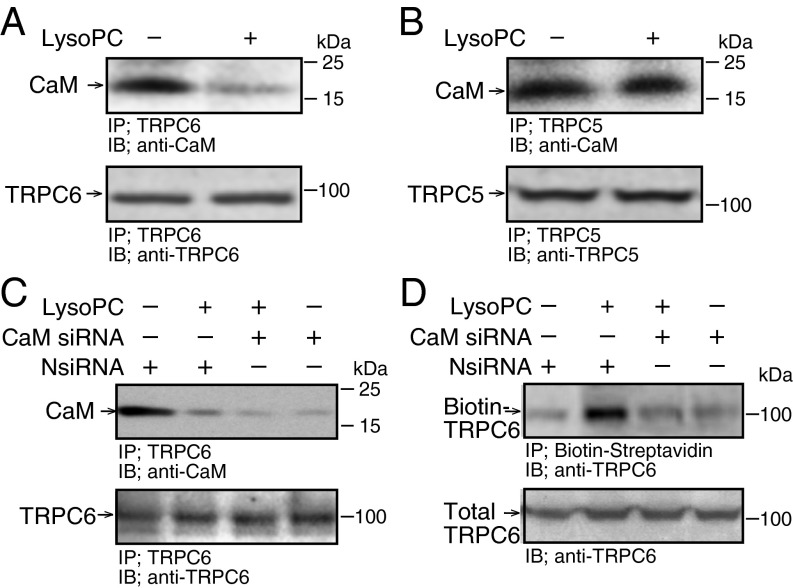
Lipid oxidation products, including lysophosphatidylcholine (lysoPC), activate canonical transient receptor potential 6 (TRPC6) channels leading to inhibition of endothelial cell (EC) migration in vitro and delayed EC healing of arterial injuries in vivo. The precise mechanism through which lysoPC activates TRPC6 channels is not known, but calmodulin (CaM) contributes to the regulation of TRPC channels. Using site-directed mutagenesis, cDNAs were generated in which Tyr(99) or Tyr(138) of CaM was replaced with Phe, generating mutant CaM, Phe(99)-CaM, or Phe(138)-CaM, respectively. In ECs transiently transfected with pcDNA3.1-myc-His-Phe(99)-CaM, but not in ECs transfected with pcDNA3.1-myc-His-Phe(138)-CaM, the lysoPC-induced TRPC6-CaM dissociation and TRPC6 externalization was disrupted. Also, the lysoPC-induced increase in intracellular calcium concentration was inhibited in ECs transiently transfected with pcDNA3.1-myc-His-Phe(99)-CaM. Blocking phosphorylation of CaM at Tyr(99) also reduced CaM association with the p85 subunit and subsequent activation of phosphatidylinositol 3-kinase (PI3K). This prevented the increase in phosphatidylinositol (3,4,5)-trisphosphate (PIP3) and the translocation of TRPC6 to the cell membrane and reduced the inhibition of EC migration by lysoPC. These findings suggest that lysoPC induces CaM phosphorylation at Tyr(99) by a Src family kinase and that phosphorylated CaM activates PI3K to produce PIP3, which promotes TRPC6 translocation to the cell membrane.

Keywords: PI3 kinase; TRPC6; calmodulin; endothelial.


Conflict of interest statement

The authors declare no conflict of interest.

Figures

Fig. 1.
Fig. 1.
LysoPC induces TRPC6-CaM dissociation but down-regulation of CaM reduces TRPC6 externalization. (A and B) BAECs were incubated with lysoPC (12.5 µM) for 15 min. TRPC6 or TRPC5 was immunoprecipitated and associated CaM identified by immunoblot analysis. In aliquots removed after immunoprecipitation, total TRPC6 or TRPC5 was determined (n = 4). (C and D) BAECs were transiently transfected with negative control siRNA (NsiRNA) or CaM siRNA (20 nM) for 24 h, then incubated with lysoPC. TRPC6-CaM association was identified as above (n = 3) or TRPC6 externalization was determined by biotinylation assay (n = 4).
Fig. S1.
Fig. S1.
CaM siRNA decreases CaM protein level. BAECs were transiently transfected with control siRNA (NsiRNA) or CaM siRNA (20 nM) for 24 h. After 48 h CaM was identified by immunoblot analysis. Actin served as loading control (n = 3). Transient transfection of BAECs with CaM small interfering RNA (siRNA), decreased CaM expression to 20 ± 3% of control (n = 3, P < 0.01).
Fig. 2.
Fig. 2.
LysoPC induces CaM phosphorylation at Tyr99, which is Ca2+ dependent, but not CaM phosphorylation at Tyr138, Ser81, or Thr79. (A–F) BAECs were incubated with lysoPC (12.5 µM) for 15 min. (A–C) Phospho-CaM was identified by immunoblot analysis. Actin served as loading control. (A) Phospho-CaM(Tyr99) was identified (n = 5). (B) Phospho-CaM(Tyr138) was identified (n = 3). Epidermal growth factor (EGF, 100 nM) for 30 min served as a positive control. (C) Phospho-CaM(Ser81/Thr79) was identified (n = 4). (D) BAECs preincubated with BAPTA/AM (25 µM) for 30 min before adding lysoPC. Phospho-CaM(Tyr99) was detected by immunoblot analysis (n = 3). (Lines indicate lanes rearranged from same gel.) (E and F) BAECs were incubated with BAPTA/AM (300 µM) for 30 min before adding lysoPC. (E) Phospho-CaM was detected by immunoblot analysis (n = 3). (F) TRPC6 externalization was determined by biotinylation assay (n = 3).
Fig. S2.
Fig. S2.
Src family tyrosine kinase inhibitor, PP2, blocks lysoPC-induced CaM phosphorylation at Tyr99, CaM dissociation from TRPC6, TRPC6 externalization, and preserved EC migration. BAECs were pretreated with PP2 (2 µM) for 1 h before incubation with lysoPC (12.5 µM). (A) Phospho-CaM(Tyr99) was identified by immunoblot analysis (n = 4). LysoPC-induced CaM phosphorylation at Tyr99 was inhibited (n = 4, P < 0.01 compared with no pretreatment). Actin served as loading control (n = 4). (B) TRPC6 was immunoprecipitated and associated CaM identified by immunoblot analysis. In aliquots removed after immunoprecipitation, total TRPC6 was determined (n = 4, P < 0.01 compared with no pretreatment). (Lines indicate lanes rearranged from same gel.) (C) TRPC6 externalization was determined by biotinylation assay. Total TRPC6 was assessed by immunoblot analysis in aliquots removed before incubation with streptavidin-agarose beads (n = 3). (D) Migration was assessed after 24 h in the presence or absence of lysoPC (12.5 µM) and results are represented as mean ± SD (n = 3, *P < 0.001 compared with control and **P < 0.001 compared with lysoPC).
Fig. S3.
Fig. S3.
LysoPC induces similar levels of CaM phosphorylation of CaM in WT and TRPC6−/− MAECs. WT or TRPC6−/− MAECs were incubated with lysoPC (10 µM for 15 min) and phospho-CaM(Tyr99) was detected by immunoblot analysis. Actin served as loading control (n = 3).
Fig. S4.
Fig. S4.
Expression of mutant CaM. (AC) BAECs were transiently transfected with pcDNA3.1-myc-His-WT-CaM or pcDNA3.1-myc-His-Phe138-CaM for 24 h. (A and B) Overexpression was confirmed after 48 h by immunoblot analysis with anti-Myc Tag antibody. Actin served as loading control (n = 3). (C) At 24 h, BAECs were incubated with lysoPC (12.5 µM) for 15 min. Phospho-CaM(Tyr99) was detected by immunoblot analysis. Actin served as loading control (n = 4). LysoPC increased CaM phosphorylation at Tyr99 1.82 ± 0.2-fold over control in BAECs overexpressing WT-CaM (P < 0.01), but not in BAECs overexpressing Phe99-CaM (n = 4). (D and E) Human endothelial cells (EA.hy926) were transiently transfected for 24 h with cDNA for WT-CaM or Phe99-CaM. (D) After 48 h, overexpression was verified by immunoblot analysis using anti-Myc tag antibody, and actin served as the loading control (n = 3). (E) Ea.hy926 cells were incubated with lysoPC (12.5 µM) for 15 min. TRPC6 externalization was determined by biotinylation assay and total TRPC6, by immunoblot analysis in aliquots removed before incubation with streptavidin-agarose beads (n = 3).
Fig. 3.
Fig. 3.
In BAECs expressing CaM mutated at Tyr99, lysoPC fails to induce TRPC6-CaM dissociation, TRPC6 tyrosine phosphorylation, and TRPC6 externalization. (AD) BAECs were transiently transfected for 24 h with pcDNA3.1-myc-His-WT-CaM, pcDNA3.1-myc-His-Phe99-CaM, or pcDNA3.1-myc-His-Phe138-CaM. At 24 h, BAECs were incubated with lysoPC (12.5 µM) for 15 min. (A) Myc-conjugated CaM was immunoprecipitated and associated TRPC6 was detected by immunoblot analysis (n = 4). (Lines indicate lanes rearranged from same gel.) (B) TRPC6 was immunoprecipitated, then immunoblot analysis using antiphosphotyrosine or anti-TRPC6 antibody identified tyrosine-phosphorylated TRPC6 or total TRPC6 (n = 4). (C and D) TRPC6 externalization was determined by biotinylation assay (n = 4).
Fig. 4.
Fig. 4.
In BAECs overexpressing Phe99-CaM, lysoPC does not increase [Ca2+]i. (AF) BAECs were transiently transfected with a plasmid containing empty vector or vector with WT-CaM, Phe99-CaM, or TRPC6, or a combination of plasmids as indicated for 16 h, made quiescent for 8 h, then loaded with fura 2-AM. After adjusting the baseline, lysoPC (12.5 µM) was added (arrow) and relative change of [Ca2+]i measured. A representative tracing of n = 8 cells is shown. (G) The mean ± SD of [Ca2+]i changes are depicted in graphic form (n = 8 measurements per condition). The change in [Ca2+]i was calculated as peak fluorescence ratio minus baseline ratio divided by baseline ratio (*P < 0.001 compared with overexpression of WT-CaM, P < 0.002 compared with vector, **P < 0.001 compared with overexpression of TRPC6 and WT-CaM).
Fig. S5.
Fig. S5.
Expression of mutant CaM and TRPC6. (A and B) BAECs were transiently transfected with a plasmid containing empty vector or vector with WT-CaM, Phe99-CaM, or TRPC6 or a combination of plasmids for 16 h. After 24 h, immunoblot analysis was performed using anti-Myc tag antibody (A, n = 3) or anti-TRPC6 antibody (B, n = 3). Actin served as loading control.
Fig. S6.
Fig. S6.
Inhibition of CaM kinase II or Pyk2 do not block lysoPC-induced TRPC6 externalization. (A) BAECs were pretreated with a CaM kinase II inhibitor, autocamtide 2-related inhibitory peptide (AIP, 10 µM) for 1 h before incubation with lysoPC (12.5 µM) for 15 min. TRPC6 externalization was determined by biotinylation assay and total TRPC6 by immunoblot analysis (n = 4). (B and C) BAECs were transiently transfected with control siRNA (NsiRNA) or Pyk2 siRNA (20 nM) for 24 h. (B) After 48 h, Pyk2 was identified by immunoblot analysis. Actin served as loading control (n = 3). (C) After incubation with lysoPC, TRPC6 externalization was detected by biotinylation assay and total TRPC6, by immunoblot analysis (n = 3).
Fig. 5.
Fig. 5.
LysoPC induces PIP3 production and PIP3-TRPC6 colocalization. (A and B) BAECs were transiently transfected with NsiRNA, p110α siRNA, or p85α siRNA, then incubated with lysoPC (12.5 µM) for 15 min. TRPC6 externalization was detected by biotinylation assay (n = 4). (C and D) BAECs were pretreated with LY294002 (20 µM) for 1 h, then lysoPC. (C) TRPC6 externalization was determined by biotinylation assay (n = 4). (D) TRPC6 was immunoprecipitated, and associated CaM or total TRPC6 was identified by immunoblot analysis (n = 3). (E) Confocal immunofluorescence microscopy identified PIP3 (green), TRPC6 (red), and PIP3-TRPC6 colocalization (yellow). Representative images of three experiments are shown. Columns 1, 2, 4, 5 show 40× magnification; column 3 shows 100× magnification. (Scale bar, 100 µm.) (F) PIP3 production was measured by ELISA and expressed as mean ± SD (n = 4, *P < 0.001 compared with control and **P < 0.02 compared with lysoPC alone). (G) WT or TRPC6−/− MAECs were incubated with lysoPC (10 µM). PIP3 production was measured by ELISA and expressed as mean ± SD (n = 3, *P < 0.01 compared with WT and **P < 0.01 compared with TRPC6−/−).
Fig. S7.
Fig. S7.
The p110α or p85α subunit of PI3K is effectively reduced by siRNA. (A and B) BAECs were transiently transfected for 24 h with control siRNA (NsiRNA 20 nM), p110α siRNA (10 nM), or p85α siRNA (20 nM). After 48 h, the p110α or p85α subunit of PI3K was identified by immunoblot analysis. Actin served as loading control (n = 3).
Fig. S8.
Fig. S8.
PIP3 production in WT or TRPC6−/− MAECs. WT or TRPC6−/− MAECs were incubated with lysoPC (10 µM) for 15 min. PIP3 was identified by confocal immunofluorescence microscopy (n = 3). Representative images are shown. Columns 1 and 2 show 40× magnification; column 3, 100× magnification. (Scale bar, 100 µm.)
Fig. 6.
Fig. 6.
In BAECs overexpressing Phe99-CaM, lysoPC does not induce CaM-PI3K association, PIP3 production, PIP3-TRPC6 colocalization, or inhibit EC migration. (A) BAECs were incubated with lysoPC (12.5 µM) for 15 min. The p85α subunit of PI3K was immunoprecipitated, and associated phospho-CaM(Tyr99), CaM, or total p85α subunit identified by immunoblot analysis (n = 4). (B–F) BAECs were transiently transfected to overexpress WT-CaM or Phe99-CaM. (B–D) BAECs were incubated with lysoPC. (B) The p85α subunit of PI3K was immunoprecipitated, and associated CaM or total p85α subunit was identified by immunoblot analysis (n = 4). (C) Confocal immunofluorescence microscopy was used to identify PIP3 (green), TRPC6 (red), or PIP3-TRPC6 colocalization (yellow). Representative images of three experiments are shown. Columns 1, 2, 3, 5 show 40× magnification; column 4 shows 100× magnification. (Scale bar, 100 µm.) (D) PIP3 production was measured by ELISA (n = 3). Results are represented as mean ± SD (*P < 0.01 compared with WT-CaM control; **P < 0.001 compared with WT-CaM incubated with lysoPC). (E) Migration was assessed after 24 h in the presence or absence of lysoPC (12.5 µM). Arrow identifies the starting line of migration. (Upper) Representative images of three experiments are shown 40× magnification. (Scale bar, 100 µm.) (Lower) Migration represented as mean ± SD (n = 3, *P < 0.001 compared with WT-CaM control, **P < 0.001 compared with Phe99-CaM control, and P < 0.001 compared with WT-CaM with lysoPC).
Fig. 7.
Fig. 7.
Model of events following EC exposure to lysoPC. LysoPC activates a Src family kinase that phosphorylates (P) CaM at Tyr99. Phosphorylated CaM dissociates from TRPC6. TRPC6 is phosphorylated. Phosphorylated CaM binds to the p85α subunit of PI3K, activating it. The increased PIP3 in the plasma membrane (PM) serves to anchor TRPC6 there. When CaM is mutated at Tyr99 to Phe99, this sequence of events is disrupted. LysoPC might initiate these events through receptor activation or release of arachidonate and activation of arachidonate-regulated Ca2+ channels. Neither the identity of the receptor (R) nor the identity of the channel activated to allow entry of the initial local Ca2+ trigger are known (brown colored).
Fig. S9.
Fig. S9.
TRPC6 externalization is not induced by saponin or blocked by membrane microviscosity stabilization or Gi-protein inhibition. (A) BAECs were incubated with the membrane detergent, saponin (0.005%), for 20 min. Incubation with lysoPC (12.5 µM) for 15 min served as a positive control. Tyrosine phosphorylation of TRPC6 was detected by immunoprecipitation of TRPC6, followed by immunoblot analysis using antiphosphotyrosine antibody. In aliquots removed after immunoprecipitation, total TRPC6 was determined by immunoblot analysis (n = 3). (B and C) BAECs were pretreated with (B) α-tocopherol (50 µM) overnight or (C) pertussis toxin (PTx, 1 μg/mL) for 24 h. LysoPC (12.5 µM) was added for 15–30 min, and TRPC6 externalization was determined by biotinylation assay. Total TRPC6 was assessed by immunoblot analysis of aliquots removed before incubation with streptavidin-agarose beads (n = 3).

Similar articles

Cited by

References

    1. Murugesan G, Fox PL. Role of lysophosphatidylcholine in the inhibition of endothelial cell motility by oxidized low density lipoprotein. J Clin Invest. 1996;97(12):2736–2744. - PMC - PubMed
    1. Tran POT, Hinman LE, Unger GM, Sammak PJ. A wound-induced [Ca2+]i increase and its transcriptional activation of immediate early genes is important in the regulation of motility. Exp Cell Res. 1999;246(2):319–326. - PubMed
    1. Chaudhuri P, et al. Elucidation of a TRPC6-TRPC5 channel cascade that restricts endothelial cell movement. Mol Biol Cell. 2008;19(8):3203–3211. - PMC - PubMed
    1. Chaudhuri P, Colles SM, Damron DS, Graham LM. Lysophosphatidylcholine inhibits endothelial cell migration by increasing intracellular calcium and activating calpain. Arterioscler Thromb Vasc Biol. 2003;23(2):218–223. - PubMed
    1. Rosenbaum MA, Chaudhuri P, Graham LM. Hypercholesterolemia inhibits re-endothelialization of arterial injuries by TRPC channel activation. J Vasc Surg. 2015;62(4):1040–1047.e2. - PMC - PubMed

Publication types

MeSH terms