. 2016 Jan 29:7:3.
doi: 10.3389/fphar.2016.00003. eCollection 2016.

Anticancer Activity of Garcinia morella on T-Cell Murine Lymphoma Via Apoptotic Induction

Affiliations

Anticancer Activity of Garcinia morella on T-Cell Murine Lymphoma Via Apoptotic Induction

Bhaswati Choudhury et al. Front Pharmacol. .

Abstract

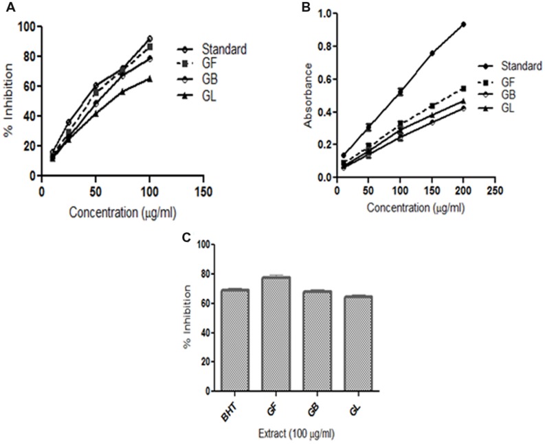
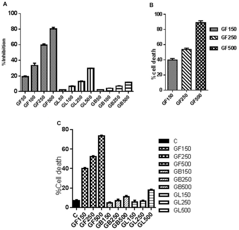
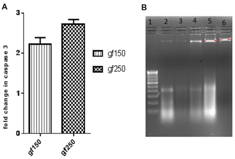
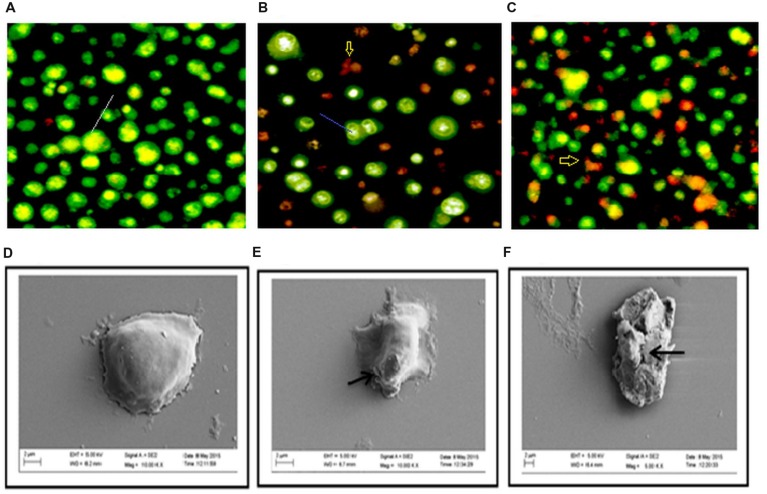
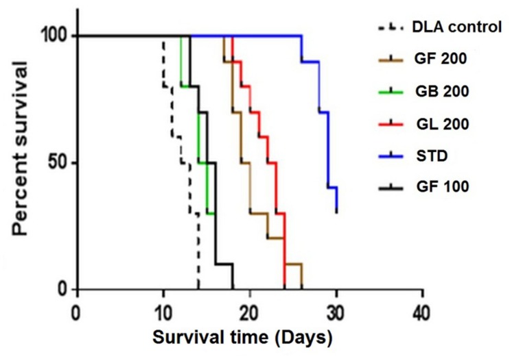
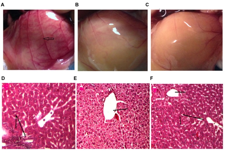
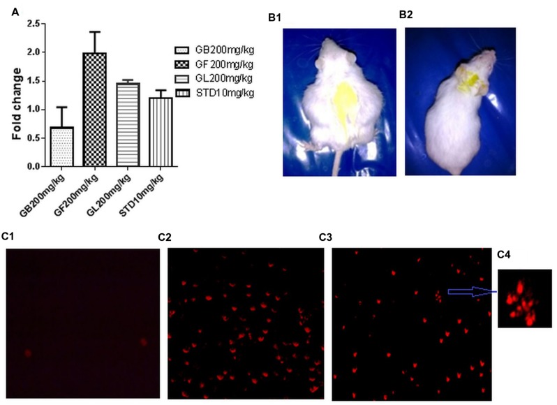
Traditional knowledge (TK) based medicines have gained worldwide attention and presently the scientific community is focussing on proper pharmacological validation and identification of lead compounds for the treatment of various diseases. The North East region of India is the home of valuable traditional herbal remedies. Garcinia morella Desr. (Guttiferae) is one such medicinal plant used by traditional healers for the treatment of inflammatory disorders. The present study was aimed to evaluate the antioxidant and anticancer activity of methanol extracts of the leaf, bark and fruit of G. morella (GM) in different in vitro and in vivo experimental conditions. The results of this study showed that GM methanol extracts possessed in vitro antioxidant and anticancer properties, where the fruit extract (GF) showed maximum activity. The anticancer activity was further confirmed by the results of in vivo administration of GF (200 mg/kg) for ten days to Dalton's lymphoma (DLA) induced mice. GF extract significantly increased the mean survival time (MST) of the animals, decreased the tumor volume and restored the hematological and biochemical parameters. The present study for the first time reported the anticancer property of GF on DLA. Further from the experiments conducted to elucidate the mechanism of action of GF on DLA, it can be concluded that GF exerts its anticancer effect through induction of caspases and DNA fragmentation that ultimately leads to apoptosis. However, further experimentation is required to elucidate the active principle and validate these findings in various in vivo settings.

Keywords: Dalton’s lymphoma; antioxidant; apoptosis; caspase3; cytotoxicity.


Figures

FIGURE 1
FIGURE 1
In vitro antioxidant assays (A) DPPH radical scavenging activity (B) Reducing power activity (C) Lipid peroxidation inhibition activity of methanol extracts of G. morella fruit (GF), bark (GB), leaf (GL), and standard BHT.
FIGURE 2
FIGURE 2
In vitro cell viability of DLA cells monitored by (A) MTT assay and (C) FACS analysis after 3 h treatment of DLA cells with different concentrations of GF, GB, and GL and (B) Trypan blue dye exclusion assay of DLA cells on treatment with GF at different concentrations for 3 h. Data are shown as % cell death for FACS and trypan blue assay and % inhibition of cell proliferation for MTT assay. % cell death and % inhibition of cell proliferation are calculated by comparing with the untreated control group. All the results are expressed in mean ± SD (n = 3).
FIGURE 3
FIGURE 3
In vitro apoptotic effect of GF on DLA cells was determined by (A) fold change in level of caspase3 of DLA cells on treatment with GF (150 and 250 μg/ml) for 3 h. Fold change is calculated by comparing with untreated DLA cells. All the results are expressed in mean ± SD (n = 3). (B) DNA fragmentation in different doses of GF treated DLA cells. 10 μg DNA from each treatment groups and untreated group was loaded in each lane and subjected to1.8% agarose gel electrophoresis followed by detection of EtBr stained DNA bands in UV transilluminator. The photograph is a representative of three repeats. Here lane1:200 bp DNA ladder, lane2, 3, 4, and 5 represents DNA of DLA cells treated with 50, 100, 150, and 200 μg/ml GF for 3 h. lane6: DNA of untreated DLA cells.
FIGURE 4
FIGURE 4
Changes in morphology of DLA cell on treatment with GF was determine by staining untreated and GF treated DLA with AO/EtBr (A–C). Here (A) is the untreated DLA cells (B) DLA cells treated with 150 μg/ml GF and (C) DLA cells treated with 250 μg/ml GF for 3 h. The white arrows are pointing to normal live cells, blue arrows are directing toward cells having shrinked nucleus representing early apoptosis and red arrows are pointing toward cells containing yellowish orange nuclei representing apoptotic cells. (D–F) Scanning electron microscope images of DLA cells afters treatment with GF 150 and 250 μg/ml for 3 h. (D) Control DLA cells. (E) DLA cells treated with GF 150 μg/ml and (F) DLA cells treated with GF 250 μg/ml for 3 h. Untreated DLA are observed to have smooth surface but the DLA cells treated with GF extract are shown have many ruptures on the surface.
FIGURE 5
FIGURE 5
Effect of the G. morella extracts on longevity of DLA induced mice is represented by Kaplan–Meir curve.
FIGURE 6
FIGURE 6
Effect of drug treatment on neo vascularisation (A–C) and liver histology (D–E). The figures (A–C) represent the inner peritoneum lining of untreated, standard treated and GF 200 mg/kg treated DLA induced animals, respectively. The blue arrow is indicating new vasculature in the peritoneum of untreated DLA induced mice. The GF treated and Standard treated animals doesn’t show any significant angiogenesis. (D–F) is the liver histology of DLA induced untreated, standard treated and GF200 mg/kg treated animals.
FIGURE 7
FIGURE 7
(A) Fold change in in vivo DLA cell death measured by FACS analysis of DLA cells aspirated from the different in vivo experimental group after staining with PI. (B1,2) are pictures of DLA induced untreated and GF 200 mg/kg treated mice after 10 days of drug treatment. (C1–3) represents the fluorescent microscope images of DLA cells aspirated from the untreated, standard treated and GF200 mg/kg treated in vivo experimental groups after staining with PI. (C4) is a zoomed image from (C3) showing apoptotic morphology of GF 200 mg/kg treated group.

Similar articles

Cited by

References

    1. Amin A., Gali-Muhtasib H., Ocker M., Schneider-Stock R. (2009). Overview of major classes of plant-derived anticancer drugs. Int. J. Biomed. Res. 5 1–11. - PMC - PubMed
    1. Anand P., Kunnumakara A. B., Sundaram C., Harikumar K. B., Tharakan S. T., Lai O. S., et al. (2008). Cancer is a preventable disease that requires major lifestyle changes. Pharm. Res. 25 2097–2116. 10.1007/s11095-008-9661-9 - DOI - PMC - PubMed
    1. Anjana A., Ahamed K. F., Ravichandiran V., Sumithra M., Anbu J. (2014). Anticancer activity of Sargassum wightii Greville on Dalton’s ascitic lymphoma. Chin. J. Nat. Med. 12 114–120. 10.1016/S1875-5364(14)60018-2 - DOI - PubMed
    1. Augustine B. B., Dash S., Lahkar M., Sarma U., Samudrala P. K., Thomas J. M. (2014). Leucas aspera inhibits the Dalton’s ascitic lymphoma in Swiss albino mice: a preliminary study exploring possible mechanism of action. Pharmacogn. Mag. 10 118–124. 10.4103/0973-1296.131022 - DOI - PMC - PubMed
    1. Badmus J. A., Adedosu T. O., Fatoki J. O., Adegbite V. A., Adaramoye O. A., Odunola O. A. (2011). Lipid peroxidation inhibition and antiradical activities of some leaf fractions of Mangifera indica. Acta Pol. Pharm. 68 23–29. - PubMed

LinkOut - more resources