. 2016 Jan 29:7:26.
doi: 10.3389/fmicb.2016.00026. eCollection 2016.

High-Throughput Isolation of Giant Viruses in Liquid Medium Using Automated Flow Cytometry and Fluorescence Staining

Affiliations

High-Throughput Isolation of Giant Viruses in Liquid Medium Using Automated Flow Cytometry and Fluorescence Staining

Jacques Y B Khalil et al. Front Microbiol. .

Abstract

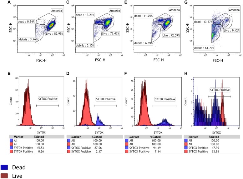
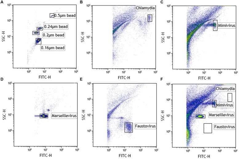
The isolation of giant viruses using amoeba co-culture is tedious and fastidious. Recently, the procedure was successfully associated with a method that detects amoebal lysis on agar plates. However, the procedure remains time-consuming and is limited to protozoa growing on agar. We present here advances for the isolation of giant viruses. A high-throughput automated method based on flow cytometry and fluorescent staining was used to detect the presence of giant viruses in liquid medium. Development was carried out with the Acanthamoeba polyphaga strain widely used in past and current co-culture experiments. The proof of concept was validated with virus suspensions: artificially contaminated samples but also environmental samples from which viruses were previously isolated. After validating the technique, and fortuitously isolating a new Mimivirus, we automated the technique on 96-well plates and tested it on clinical and environmental samples using other protozoa. This allowed us to detect more than 10 strains of previously known species of giant viruses and seven new strains of a new virus lineage. This automated high-throughput method demonstrated significant time saving, and higher sensitivity than older techniques. It thus creates the means to isolate giant viruses at high speed.

Keywords: automated system; flow cytometry; fluorescence staining; gating strategy; giant viruses; high-throughput; protozoa.


Figures

FIGURE 1
FIGURE 1
Detection of potential amoebal infection. A living amoebal population was used as a negative control with Sytox staining. Three gates were designed in the FSC-SSC plot, potentially corresponding to live cells, dead cells and debris. At 0 h the negative control and sample showed the same gated populations (A,B). After 24 h of culture, a physiologic increase in dead cells was observed for both the negative control and test samples (C,D). The appearance of a large population of debris confirmed a potential infection after 48 h of culture and a larger part of dead cells in the tested samples than in the negative control showing no substantial changes in the gated populations (E,F representing the negative control panel after 48 h and, G,H representing the sample containing Mimivirus after 48 h).
FIGURE 2
FIGURE 2
Cytometric analysis of amoebal pathogens: giant viruses and chlamydiae. (A) Using Megamix+ SSC beads (0.16-, 0.2-, 0.24-, and 0.5-μm fluorescent beads), LSR fortessa was easily set up for SSC, and a 520 nm Fluorescent channel (for SYBR DNA virus labeling) was used for virus range analysis. Fluorescence threshold was set to limit background noise of the instrument. Each microorganism was first analyzed alone to clearly define virus gates [(B) chlamydia, (C) Mimivirus, (D) Marseillevirus, and (E) Faustovirus]. Thanks to the wide resolution of LSR fortessa in SSC and the clear DNA content between the different biological events, four different gates were created. Due to the high concentration of virus in the supernatant after amoebal burst, a virus was considered present if its concentration was more than 103 virus/μl (counting beads). (F) Viral mixture of Mimivirus and Marseillevirus, detected in the same sample.

References

    1. Abrahão J. S., Dornas F. P., Silva L. C. F., Almeida G. M., Boratto P. V. M., Colson P., et al. (2014). Acanthamoeba polyphaga mimivirus and other giant viruses: an open field to outstanding discoveries. Virol. J. 11:120 10.1186/1743-422X-11-120 - DOI - PMC - PubMed
    1. Avery S. V. S., Harwood J. L. J., Lloyd D. D. (1995). Quantification and characterization of phagocytosis in the soil amoeba Acanthamoeba castellanii by flow cytometry. Appl. Environ. Microbiol. 61 1124–1132. - PMC - PubMed
    1. Boughalmi M., Saadi H., Pagnier I., Colson P., Fournous G., Raoult D., et al. (2012). High-throughput isolation of giant viruses of the Mimiviridae and Marseilleviridae families in the Tunisian environment. Environ. Microbiol. 15 2000–2007. 10.1111/1462-2920.12068 - DOI - PubMed
    1. Boyer M., Yutin N., Pagnier I., Barrassi L., Fournous G., Espinosa L., et al. (2009). Giant Marseillevirus highlights the role of amoebae as a melting pot in emergence of chimeric microorganisms. Proc. Natl. Acad. Sci. U.S.A. 106 21848–21853. 10.1073/pnas.0911354106 - DOI - PMC - PubMed
    1. Brussaard C. P. D. (2004). Optimization of procedures for counting viruses by flow cytometry. Appl. Environ. Microbiol. 70 1506–1513. 10.1128/AEM.70.3.1506-1513.2004 - DOI - PMC - PubMed

LinkOut - more resources