. 2016 Jan 13;10(1):014106.
doi: 10.1063/1.4940041. eCollection 2016 Jan.

Human induced pluripotent stem cells derived endothelial cells mimicking vascular inflammatory response under flow

Affiliations

Human induced pluripotent stem cells derived endothelial cells mimicking vascular inflammatory response under flow

Li Wang et al. Biomicrofluidics. .

Abstract

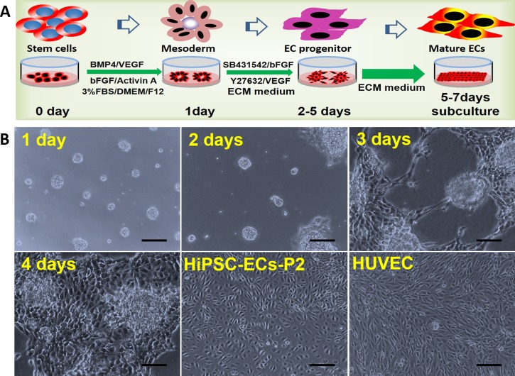
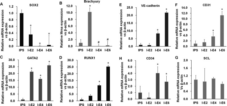
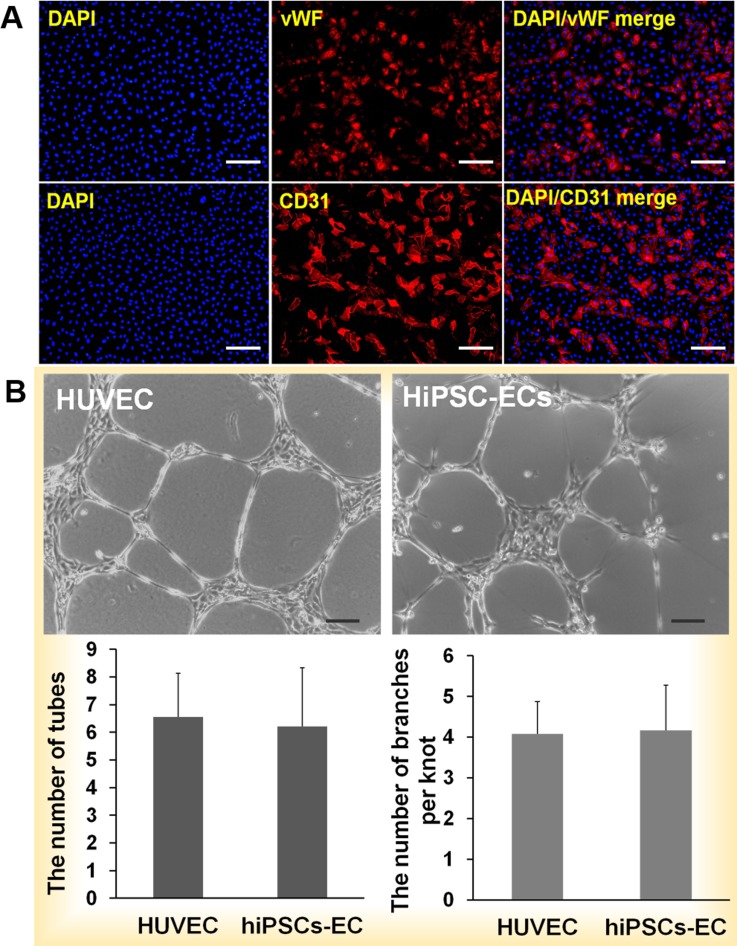
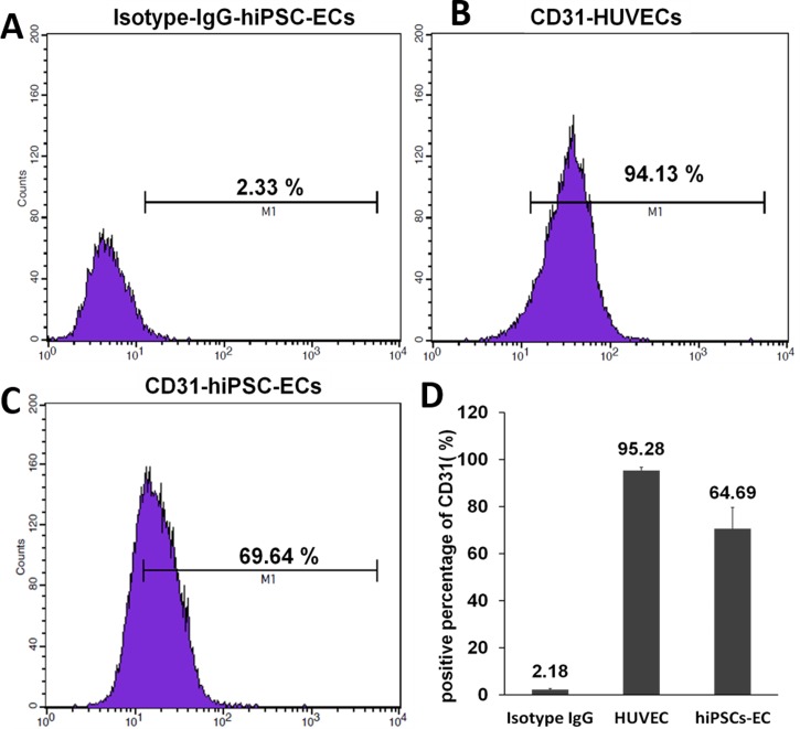
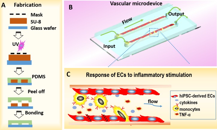
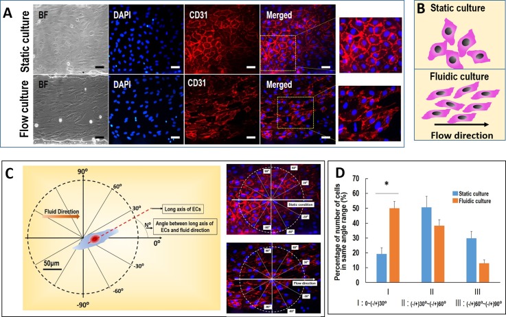
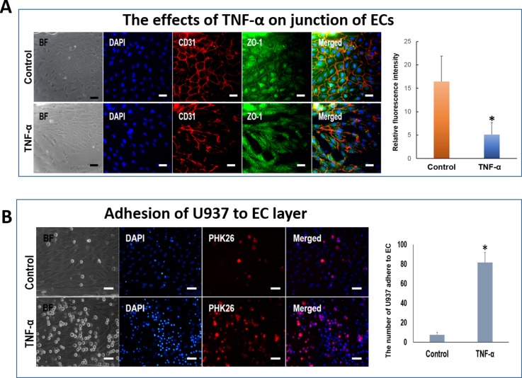
Endothelial cells (ECs) have great potential in vascular diseases research and regenerative medicine. Autologous human ECs are difficult to acquire in sufficient numbers in vitro, and human induced pluripotent stem cells (iPSCs) offer unique opportunity to generate ECs for these purposes. In this work, we present a new and efficient method to simply differentiate human iPSCs into functional ECs, which can respond to physiological level of flow and inflammatory stimulation on a fabricated microdevice. The endothelial-like cells were differentiated from human iPSCs within only one week, according to the inducing development principle. The expression of endothelial progenitor and endothelial marker genes (GATA2, RUNX1, CD34, and CD31) increased on the second and fourth days after the initial inducing process. The differentiated ECs exhibited strong expression of cells-specific markers (CD31 and von Willebrand factor antibody), similar to that present in human umbilical vein endothelial cells. In addition, the hiPSC derived ECs were able to form tubular structure and respond to vascular-like flow generated on a microdevice. Furthermore, the human induced pluripotent stem cell-endothelial cells (hiPSC-ECs) pretreated with tumor necrosis factor (TNF-α) were susceptible to adhesion to human monocyte line U937 under flow condition, indicating the feasibility of this hiPSCs derived microsystem for mimicking the inflammatory response of endothelial cells under physiological and pathological process.


Figures

FIG. 1.
FIG. 1.
Differentiation protocol and morphology of hiPSC-ECs. (a) Schematic schedule of the differentiation of hiPSCs into ECs. (b) The hiPSCs formed circular colonies on the first day. Some cells sprouted from the cell aggregates on the second day, and the sprouting branches extended gradually on the third and fourth days. The migrating cells reached 90% confluence on the fifth through seventh days. After scraping away the non-ECs, the cells derived from hiPSCs exhibited cobblestone morphology by passage 2 and appeared similar to HUVECs. P2, passage 2. Scale bar = 100 μm.
FIG. 2.
FIG. 2.
Gene expression during differentiation. (a) The levels of SOX2 (pluripotency marker) decreased rapidly on the second day of culture. (b) Transcription of the T gene encoding brachyury (mesodermal marker) peaked on the second day and then decreased rapidly. (c) and (d) Transcription of the genes encoding the angioblast markers GATA2 and RUNX1 increased after 2 days. (e)–(g) Transcription of the genes encoding VE-cadherin, CD31, and CD34 started to increase on day 4. (h) The level of transcription of SCL was unchanged and that of CD34 increased slightly. I–E(n) = iPSC-ECs (days of differentiation).
FIG. 3.
FIG. 3.
Identification of hiPSC-derived ECs and tube formation. (a) Immunofluorescence analysis of CD31 and vWF expression by hiPSC-derived ECs. Most of the cells derived from hiPSCs expressed the endothelial cell-specific markers CD31 on membranes and vWF in the cytoplasm. HUVECs expressed CD31 on the outer membrane. The nuclei were counterstained with DAPI. (b) hiPSC-ECs-P1 and HUVECs-P1 were cultured on Matrigel for 24 h, and sprouting cells formed a network among the cellular aggregates. The numbers of complete tubes and branches per knot were similar to those of HUVECs. Scale bar = 100 μm.
FIG. 4.
FIG. 4.
Flow cytometric analysis of CD31 expression by hiPSC-ECs. (a) Detection using an IgG isotype matched antibody (negative control). (b) Over 90% HUVECs were CD31-postive. (c) Over 60% hiPSC-ECs were CD31-positive. (d) The average number of CD31-positive cells was calculated for three independent experiments. The average values are shown above the columns.
FIG. 5.
FIG. 5.
Schematic of microdevice and vascular inflammation. (a) The procedure of fabricating microdevice. (b) The schematic diagram of vascular inflammation on chip. The microdevice has three parts: a glass substrate layer, a layer of polydimethylsiloxane (PDMS) with channels, and a perfusion system. (c) The enlarged view of red frame in B represents the interaction between inflammatory cells and endothelial cells.
FIG. 6.
FIG. 6.
The effects of flow stress on the arrangement of hiPSC-ECs. (a) Immunofluorescence assay showing the morphology of hiPSC-ECs under physiological relevant flow stress using CD31 antibodies. The nuclei were counterstained with DAPI. (b) The schematic diagram of the arrangement of hiPSC-ECs under static culture and flow culture. The hiPSC-ECs aligned with flow direction. (c) and (d) The arrangement of hiPSC-ECs under flow stress. The cellular arrangement was defined as the angle between the cellular long axis and the flow direction. The number of cells within same angle area in unit circle area was counted according to the cell arrangement. The angular range was divide as −30° to +30°, 30° to 60°/−30° to −60°, 60° to 90°/−60° to −90°. BF: bright field. Scale bar = 50 μm.
FIG. 7.
FIG. 7.
Interaction between U937 cells and hiPSC-ECs under inflammation. (a) The hiPSC-ECs in microchannels were treated with TNF-α under low flow stress. The membrane protein CD31 and tight junction protein ZO-1 were stained by immunofluorescence assay. The nuclei were counterstained with DAPI. The histogram indicates the relative fluorescence intensity of ZO-1. (b) U937 cells adhering to the hiPSC-ECs in microchannels under inflammation conditions treated with TNF-α. Normal medium was used as the control. Right figure indicates that number of U937 cells adhering to the ECs. BF: bright field. Scale bar = 50 μm.

Similar articles

Cited by

References

    1. Carmeliet P., Nature 438, 932–936 (2005).10.1038/nature04478 - DOI - PubMed
    1. Park K. H. and Park W. J., J. Korean Med. Sci. 30, 1213–1225 (2015).10.3346/jkms.2015.30.9.1213 - DOI - PMC - PubMed
    1. Sampson U. K., Engelgau M. M., Peprah E. K., and Mensah G. A., Cardiovasc. J. Afr. 26, S56–S60 (2015).10.5830/CVJA-2015-043 - DOI - PMC - PubMed
    1. Polovina M. M. and Potpara T. S., Postgrad. Med. 126, 38–53 (2014).10.3810/pgm.2014.03.2739 - DOI - PubMed
    1. Takahashi K. and Yamanaka S., Cell 126, 663–676 (2006).10.1016/j.cell.2006.07.024 - DOI - PubMed