

Enhanced contactless dielectrophoresis enrichment and isolation platform via cell-scale microstructures
- PMID: 26858821
- PMCID: PMC4723398
- DOI: 10.1063/1.4939947
Enhanced contactless dielectrophoresis enrichment and isolation platform via cell-scale microstructures
Abstract
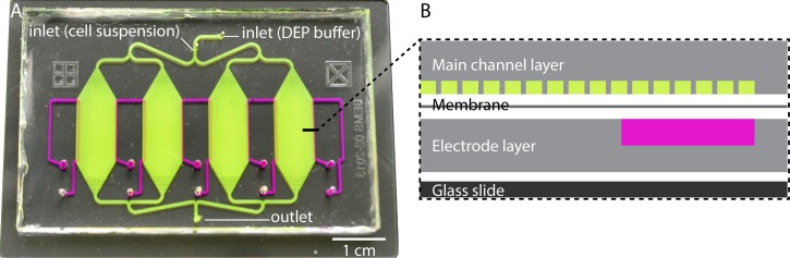
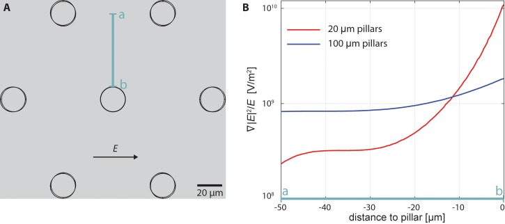
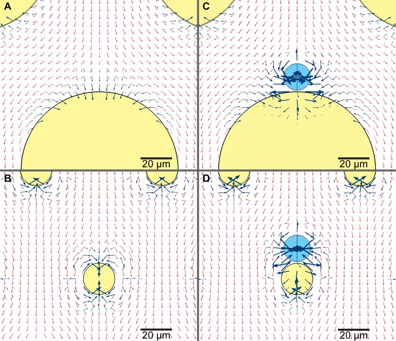
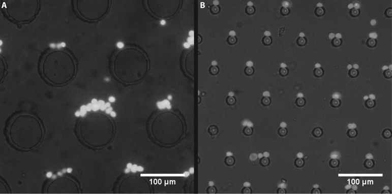
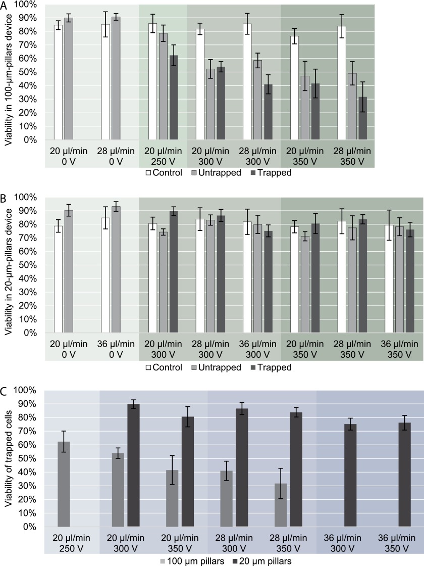
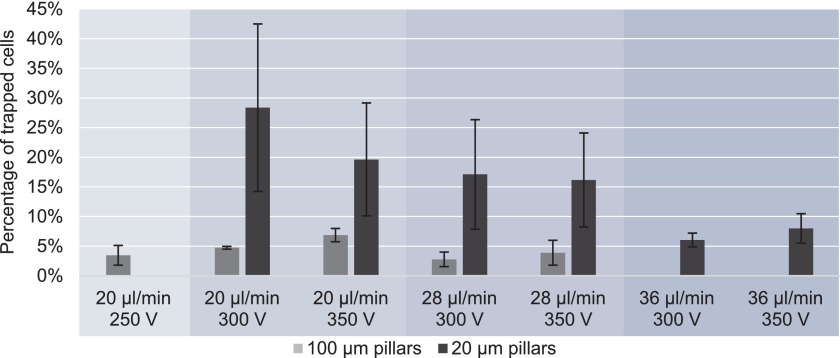
We designed a new microfluidic device that uses pillars on the same order as the diameter of a cell (20 μm) to isolate and enrich rare cell samples from background. These cell-scale microstructures improve viability, trapping efficiency, and throughput while reducing pearl chaining. The area where cells trap on each pillar is small, such that only one or two cells trap while fluid flow carries away excess cells. We employed contactless dielectrophoresis in which a thin PDMS membrane separates the cell suspension from the electrodes, improving cell viability for off-chip collection and analysis. We compared viability and trapping efficiency of a highly aggressive Mouse Ovarian Surface Epithelial (MOSE) cell line in this 20 μm pillar device to measurements in an earlier device with the same layout but pillars of 100 μm diameter. We found that MOSE cells in the new device with 20 μm pillars had higher viability at 350 VRMS, 30 kHz, and 1.2 ml/h (control 77%, untrapped 71%, trapped 81%) than in the previous generation device (untrapped 47%, trapped 42%). The new device can trap up to 6 times more cells under the same conditions. Our new device can sort cells with a high flow rate of 2.2 ml/h and throughput of a few million cells per hour while maintaining a viable population of cells for off-chip analysis. By using the device to separate subpopulations of tumor cells while maintaining their viability at large sample sizes, this technology can be used in developing personalized treatments that target the most aggressive cancerous cells.
Figures
Similar articles
-
Enhanced Particle Trap: Design and Simulation of Pillar-Based Contactless Dielectrophoresis Microfluidic Devices.Electrophoresis. 2025 Feb;46(3-4):232-239. doi: 10.1002/elps.202400110. Epub 2025 Feb 18. Electrophoresis. 2025. PMID: 39965079
-
A feasibility study for enrichment of highly aggressive cancer subpopulations by their biophysical properties via dielectrophoresis enhanced with synergistic fluid flow.Electrophoresis. 2017 Jun;38(11):1507-1514. doi: 10.1002/elps.201600530. Epub 2017 May 8. Electrophoresis. 2017. PMID: 28342274 Free PMC article.
-
Numerical evaluation and experimental validation of cross-flow microfiltration device design.Biomed Microdevices. 2019 Feb 21;21(1):21. doi: 10.1007/s10544-019-0378-9. Biomed Microdevices. 2019. PMID: 30790088
-
A pillar-based microfilter for isolation of white blood cells on elastomeric substrate.Biomicrofluidics. 2013 Jan 9;7(1):14102. doi: 10.1063/1.4774068. eCollection 2013. Biomicrofluidics. 2013. PMID: 24403994 Free PMC article.
-
Electric field-induced effects on neuronal cell biology accompanying dielectrophoretic trapping.Adv Anat Embryol Cell Biol. 2003;173:III-IX, 1-77. doi: 10.1007/978-3-642-55469-8. Adv Anat Embryol Cell Biol. 2003. PMID: 12901336 Review.
Cited by
-
Integrated dielectrophoretic and surface plasmonic platform for million-fold improvement in the detection of fluorescent events.Biomicrofluidics. 2017 Aug 22;11(4):044115. doi: 10.1063/1.5000008. eCollection 2017 Jul. Biomicrofluidics. 2017. PMID: 28868108 Free PMC article.
-
Microfluidics as efficient technology for the isolation and characterization of stem cells.EXCLI J. 2021 Feb 22;20:426-443. doi: 10.17179/excli2020-3028. eCollection 2021. EXCLI J. 2021. PMID: 33746671 Free PMC article. Review.
-
Separation of Macrophages and Fibroblasts Using Contactless Dielectrophoresis and a Novel ImageJ Macro.Bioelectricity. 2019 Mar 1;1(1):49-55. doi: 10.1089/bioe.2018.0004. Epub 2019 Mar 18. Bioelectricity. 2019. PMID: 32292890 Free PMC article.
-
Bridging the scales in high-throughput dielectrophoretic (bio-)particle separation in porous media.Sci Rep. 2018 Jul 11;8(1):10480. doi: 10.1038/s41598-018-28735-w. Sci Rep. 2018. PMID: 29993026 Free PMC article.
-
Alternative cDEP Design to Facilitate Cell Isolation for Identification by Raman Spectroscopy.Sensors (Basel). 2017 Feb 9;17(2):327. doi: 10.3390/s17020327. Sensors (Basel). 2017. PMID: 28208767 Free PMC article.
References
Grants and funding
LinkOut - more resources
Full Text Sources
Other Literature Sources