Improved Hepatocyte Engraftment After Portal Vein Occlusion in LDL Receptor-Deficient WHHL Rabbits and Lentiviral-Mediated Phenotypic Correction In Vitro
- PMID: 26858856
- PMCID: PMC4733829
- DOI: 10.3727/215517912X647136
Improved Hepatocyte Engraftment After Portal Vein Occlusion in LDL Receptor-Deficient WHHL Rabbits and Lentiviral-Mediated Phenotypic Correction In Vitro
Abstract
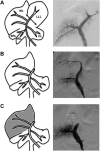
Innovative cell-based therapies are considered as alternatives to liver transplantation. Recent progress in lentivirus-mediated hepatocyte transduction has renewed interest in cell therapy for the treatment of inherited liver diseases. However, hepatocyte transplantation is still hampered by inefficient hepatocyte engraftment. We previously showed that partial portal vein embolization (PVE) improved hepatocyte engraftment in a nonhuman primate model. We developed here an ex vivo approach based on PVE and lentiviral-mediated transduction of hepatocytes from normal (New Zealand White, NZW) and Watanabe heritable hyperlipidemic (WHHL) rabbits: the large animal model of familial hypercholesterolemia type IIa (FH). FH is a life-threatening human inherited autosomal disease caused by a mutation in the low-density lipoprotein receptor (LDLR) gene, which leads to severe hypercholesterolemia and premature coronary heart disease. Rabbit hepatocytes were isolated from the resected left liver lobe, and the portal branches of the median lobes were embolized with Histoacryl® glue under radiologic guidance. NZW and WHHL hepatocytes were each labeled with Hoechst dye or transduced with lentivirus expressing GFP under the control of a liver-specific promoter (mTTR, a modified murine transthyretin promoter) and were then immediately transplanted back into donor animals. In our conditions, 65-70% of the NZW and WHHL hepatocytes were transduced. Liver repopulation after transplantation with the Hoechst-labeled hepatocytes was 3.5 ± 2%. It was 1.4 ± 0.6% after transplantation with either the transduced NZW hepatocytes or the transduced WHHL hepatocytes, which was close to that obtained with Hoechst-labeled cells, given the mean transduction efficacy. Transgene expression persisted for at least 8 weeks posttransplantation. Transduction of WHHL hepatocytes with an LDLR-encoding vector resulted in phenotypic correction in vitro as assessed by internalization of fluorescent LDL ligands. In conclusion, our results have applications for the treatment of inherited metabolic liver diseases, such as FH, by transplantation of lentivirally transduced hepatocytes.
Keywords: Familial hypercholesterolemia; Hepatocyte transplantation; Lentiviral vector; Liver; Portal vein embolization; Rabbit.
Figures


Similar articles
-
Efficient hepatocyte engraftment and long-term transgene expression after reversible portal embolization in nonhuman primates.Hepatology. 2009 Mar;49(3):950-9. doi: 10.1002/hep.22739. Hepatology. 2009. PMID: 19152424
-
Transplantation of genetically modified hepatocytes after liver preconditioning in Watanabe heritable hyperlipidemic rabbit.J Surg Res. 2018 Apr;224:23-32. doi: 10.1016/j.jss.2017.11.060. Epub 2017 Dec 22. J Surg Res. 2018. PMID: 29506845
-
Ex vivo gene therapy of familial hypercholesterolemia.Hum Gene Ther. 1992 Apr;3(2):179-222. doi: 10.1089/hum.1992.3.2-179. Hum Gene Ther. 1992. PMID: 1391038
-
Watanabe heritable hyperlipidemic rabbit. Animal model for familial hypercholesterolemia.Arteriosclerosis. 1989 Jan-Feb;9(1 Suppl):I33-8. Arteriosclerosis. 1989. PMID: 2643428 Review.
-
Cell transplantation in liver-directed gene therapy.Cell Transplant. 1993 Sep-Oct;2(5):381-400; discussion 407-10. doi: 10.1177/096368979300200504. Cell Transplant. 1993. PMID: 8162279 Review.
Cited by
-
Human hepatocyte transplantation for liver disease: current status and future perspectives.Pediatr Res. 2018 Jan;83(1-2):232-240. doi: 10.1038/pr.2017.284. Epub 2017 Dec 6. Pediatr Res. 2018. PMID: 29149103 Review.
-
Human Hepatocyte Transplantation: Three Decades of Clinical Experience and Future Perspective.Stem Cells Transl Med. 2024 Mar 15;13(3):204-218. doi: 10.1093/stcltm/szad084. Stem Cells Transl Med. 2024. PMID: 38103170 Free PMC article. Review.
References
-
- Allen K. J.; Mifsud N. A.; Williamson R.; Bertolino P.; Hardikar W. Cell-mediated rejection results in allograft loss after liver cell transplantation. Liver Transpl. 14(5):688–694; 2008. - PubMed
-
- Ambrosino G.; Varotto S.; Strom S. C.; Guariso G.; Franchin E.; Miotto D.; Caenazzo L.; Basso S.; Carraro P.; Valente M. L.; D'Amico D.; Zancan L.; D'Antiga L. Isolated hepatocyte transplantation for Crigler–Najjar syndrome type 1. Cell Transplant. 14(2–3):151–157; 2005. - PubMed
-
- Andreoletti M.; Loux N.; Vons C.; Nguyen T. H.; Lorand I.; Mahieu D.; Simon L.; Di Rico V.; Vingert B.; Chapman J.; Briand P.; Schwall R.; Hamza J.; Capron F.; Bargy F.; Franco D.; Weber A. Engraftment of autologous retrovirally transduced hepatocytes after intraportal transplantation into nonhuman primates: Implication for ex vivo gene therapy. Hum. Gene Ther. 12(2):169–179; 2001. - PubMed
-
- Attaran M.; Schneider A.; Grote C.; Zwiens C.; Flemming P.; Gratz K. F.; Jochheim A.; Bahr M. J.; Manns M. P.; Ott M. Regional and transient ischemia/reperfusion injury in the liver improves therapeutic efficacy of allogeneic intraportal hepatocyte transplantation in low-density lipoprotein receptor deficient Watanabe rabbits. J. Hepatol. 41(5):837–844; 2004. - PubMed
-
- Aurich H.; Sgodda M.; Kaltwasser P.; Vetter M.; Weise A.; Liehr T.; Brulport M.; Hengstler J. G.; Dollinger M. M.; Fleig W. E.; Christ B. Hepatocyte differentiation of mesenchymal stem cells from human adipose tissue in vitro promotes hepatic integration in vivo. Gut 58(4):570–581; 2009. - PubMed
LinkOut - more resources
Full Text Sources
Research Materials
Miscellaneous

