

doi: 10.1016/j.mmcr.2015.12.003.
eCollection 2015 Dec.
Aggressive cutaneous zygomycosis caused by Apophysomyces variabilis in an immunocompetent child
Affiliations
- PMID: 26858932
- PMCID: PMC4706566
- DOI: 10.1016/j.mmcr.2015.12.003
Item in Clipboard
Aggressive cutaneous zygomycosis caused by Apophysomyces variabilis in an immunocompetent child
Med Mycol Case Rep.
.
Abstract
A zygomycetous fungus was observed in a biopsy of a 9-year-old male. The patient was presented with severe cutaneous lesions subsequent to a traumatic car accident. Following fungal detection, antifungal treatment was prescribed but condition deteriorated rapidly and above knee amputation was done as lifesaving and to control fungal infection. Analysis of the 28 S rRNA gene (accession KT149770) aligned the isolate with members of the genus Apophysomyces and the pathogen was identified as Apophysomces variabilis.
Keywords: 28S rRNA gene; Amputation; Apophysomyces; Aseer region; Zygomycetes.
Figures
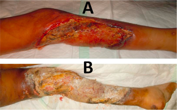
Massive ulcerated cutaneous zygomycosis lesions on the left leg of the 9 years old patient. Lesions seen after two weeks (A) and wide spread necrosis of skin and soft tissue that extended from mid-thigh down to the left ankle seen just prior to amputation (B).
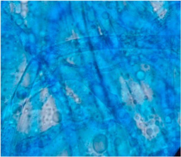
A direct microscopy micrograph of smear from debrided tissues obtained from the 9 year old male patient suffering from a subcutaneous wound infection. Note the broad hyaline multinucleated aseptate hyphae suggestive of a zygomycetous fungus.
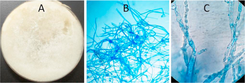
Growth of the zygomatous fungus (AB7-1) on Sabouraud dextrose agar (A) showing heavy white mold. Under the microscope the mold shows broad hyaline aseptate hyphae (B, C).
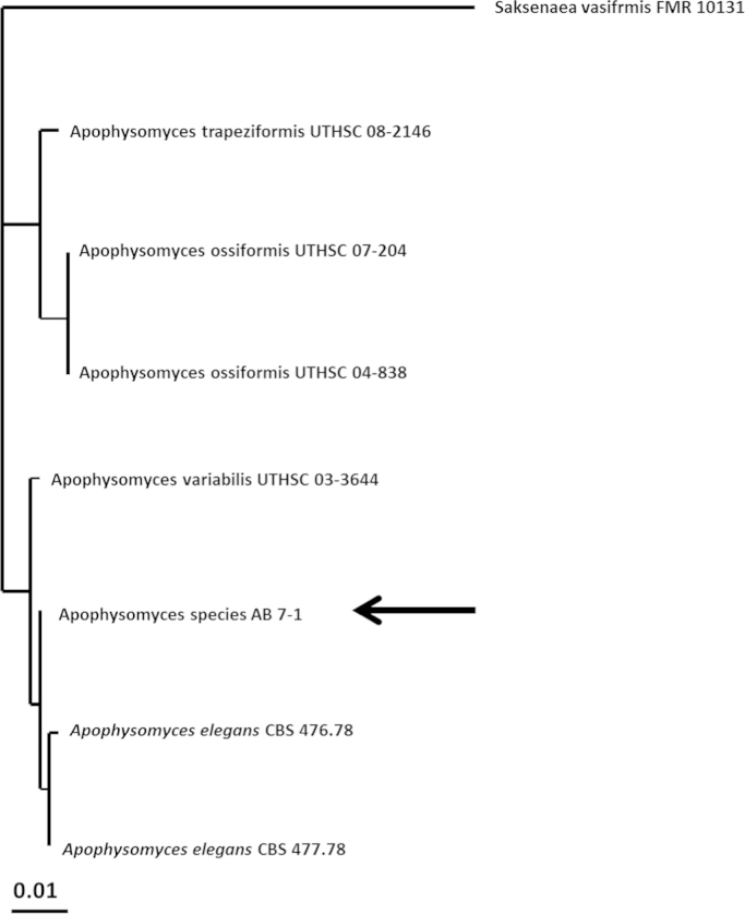
Estimate of Apophysomces phylogeny based on sequence analysis of domains D1 and D2 of the 28 S rRNA gene (accession KT149770) showing the taxonomic position of the strain AB7-1 (arrow) within members of the genus Apophysomces. The analysis shows that the strain is placed within a clade encompassing A. elegans and A. variabilis.
References
-
- Linder N., Keller N., Huri C., Kuint J., Goldshmidt-Reuven A., Barzilai A. Primary cutaneous mucormycosis in a premature infant: case report and review of the literature. Am. J. Perinatol. 1998;15:35–38. - PubMed
-
- Ruiz C.E., Arango M., Correa A.L., Lopez L.S., Restrepo A. [Necrotizing fasciitis in an immunocompetent patient caused by Apophysomyces elegans] Biomedica. 2004;24:239–251. - PubMed
-
- Weddle G., Gandy K., Bratcher D., Pahud B., Jackson M.A. Apophysomyces trapeziformis infection associated with a tornado-related injury. Pediatr. Infect. Dis. J. 2012;31:640–642. - PubMed
LinkOut - more resources
Full Text Sources
Other Literature Sources
Molecular Biology Databases
