

Perineural Injection for Treatment of Root-Signature Signs Associated with Lateralized Disk Material in Five Dogs (2009-2013)
- PMID: 26858952
- PMCID: PMC4728328
- DOI: 10.3389/fvets.2016.00001
Perineural Injection for Treatment of Root-Signature Signs Associated with Lateralized Disk Material in Five Dogs (2009-2013)
Abstract
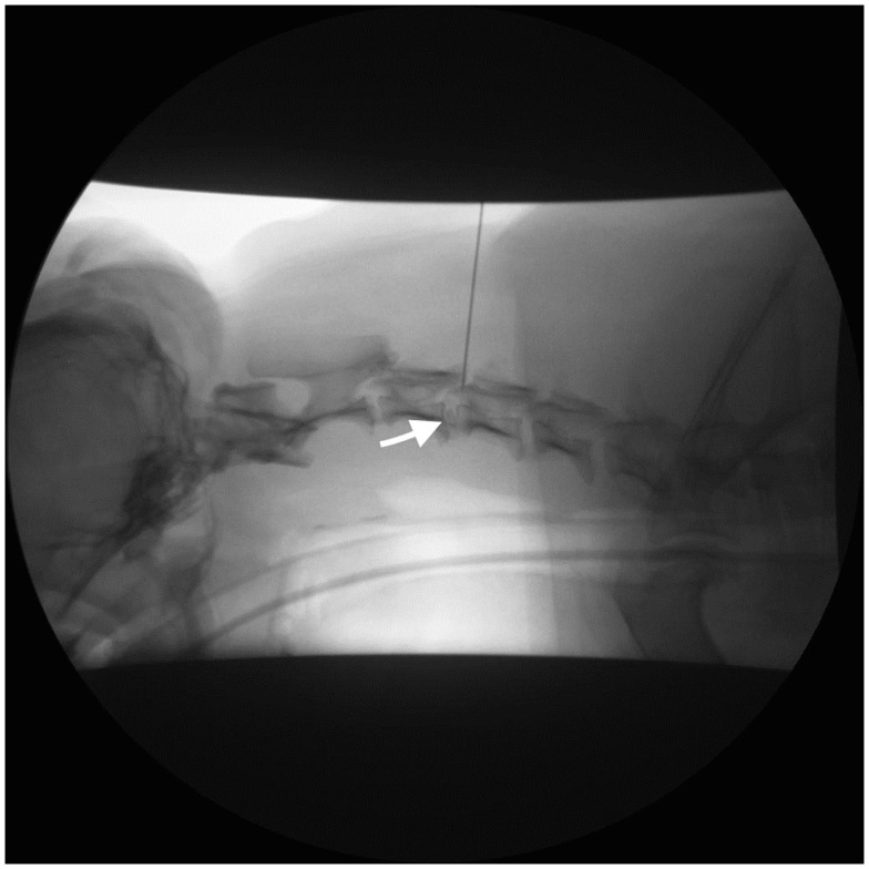
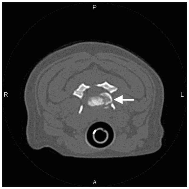
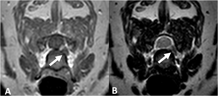
Intervertebral disk disease (IVDD) is common in dogs; cervical IVDD accounts for 13-25% of all cases. Ventral slot decompression provides access to ventral and centrally extruded or protruded disk material. However, procedures to remove dorsally or laterally displaced material are more difficult. This case series describes the use of perineural injection as a potential treatment option for dogs experiencing root-signature signs associated with lateralized disk material in the cervical spine. Five dogs underwent fluoroscopically guided perineural injection of methylprednisolone ± bupivacaine. Most patients experienced improvement in root-signature signs and remained pain free without the assistance of oral pain medication. These findings suggest the perineural injection of methylprednisolone ± bupivacaine represents a viable option for dogs with cervical lateralized disk material causing root-signature signs.
Keywords: fluoroscopy; intervertebral disk disease; lateralized disk; perineural; root-signature.
Figures
References
-
- Bagley RS, Tucker R, Harrington ML. Lateral and foraminal disk extrusion in dogs. Comp Cont Educ Pract Vet (1996) 18(7):795–804.
-
- Smith BA, Hosgood G, Kerwin SC. Ventral slot decompression for cervical intervertebral disc disease in 112 dogs. Austr Vet Pract (1997) 27(2):58–64.
-
- Fry TR, Johnson AL, Hungerford L. Surgical treatment of cervical disc herniation in ambulatory dogs, ventral decompression vs fenestration, 111 cases (1980-1988). Prog Vet Neurol (1991) 2:165–73.
-
- Seim HB, III, Prata RG. Ventral decompression for the treatment of cervical disk disease in the dog: a review of 54 cases. J Am Anim Hosp Assoc (1982) 18:233–40.
Publication types
LinkOut - more resources
Full Text Sources
Other Literature Sources