Review
. 2016 Feb 8;26(3):R125-39.
doi: 10.1016/j.cub.2015.12.032.

Fertilization Mechanisms in Flowering Plants

Affiliations
Review

Fertilization Mechanisms in Flowering Plants

Thomas Dresselhaus et al. Curr Biol. .

Abstract

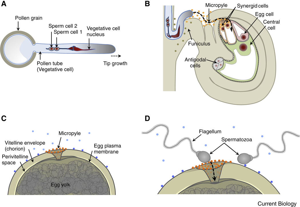
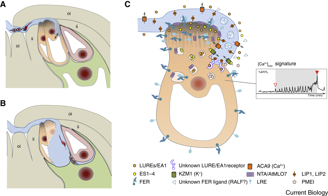
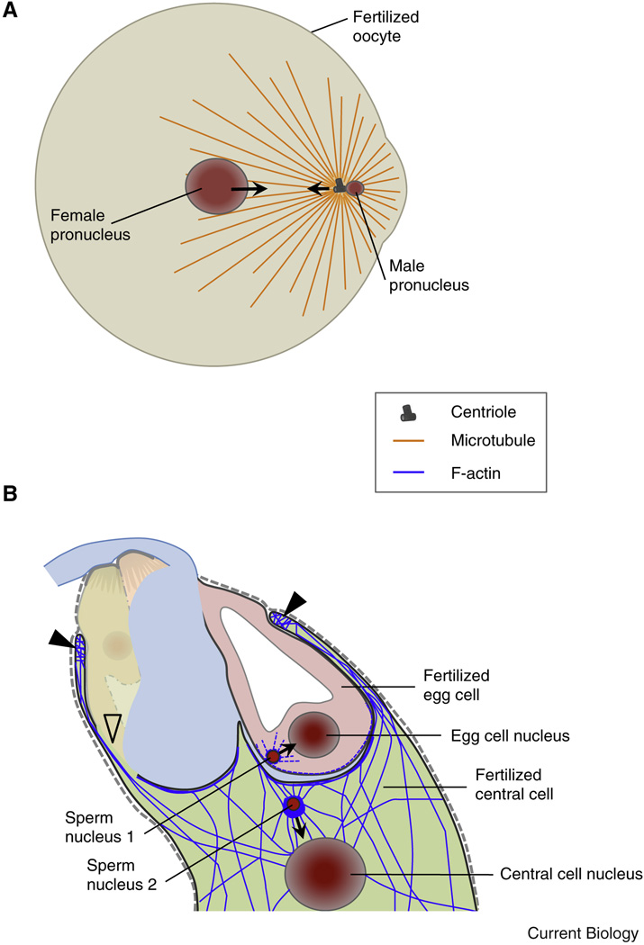
Compared with the animal kingdom, fertilization is particularly complex in flowering plants (angiosperms). Sperm cells of angiosperms have lost their motility and require transportation as a passive cargo by the pollen tube cell to the egg apparatus (egg cell and accessory synergid cells). Sperm cell release from the pollen tube occurs after intensive communication between the pollen tube cell and the receptive synergid, culminating in the lysis of both interaction partners. Following release of the two sperm cells, they interact and fuse with two dimorphic female gametes (the egg and the central cell) forming the major seed components embryo and endosperm, respectively. This process is known as double fertilization. Here, we review the current understanding of the processes of sperm cell reception, gamete interaction, their pre-fertilization activation and fusion, as well as the mechanisms plants use to prevent the fusion of egg cells with multiple sperm cells. The role of Ca(2+) is highlighted in these various processes and comparisons are drawn between fertilization mechanisms in flowering plants and other eukaryotes, including mammals.


Figures

Figure 1
Figure 1. Comparison of gamete/gametophyte morphology between the model plant Arabidopsis thaliana and the model animal Zebrafish
(A) Diagram of the haploid male gametophyte (pollen) of Arabidopsis comprising the vegetative cell (producing the growing pollen tube) and two non-motile sperm cells enclosed within the a membrane of the vegetative tube cell. The sperm cells are connected to each other and to the nucleus of the vegetative pollen tube cell forming the "male germ unit". Nuclei in red, vegetative cell membrane in blue, sperm cell membranes in black. (B) A pollen tube approaching the Arabidopsis ovule. The tube grows through the micropyle of the ovule along the funiculus towards the haploid female gametophyte that comprises the egg cell, central cell and accessory cells (synergid and antipodal cells). Secreted LURE peptides (orange dots) act as pollen tube attractants guiding the pollen tube through the micropyle. Other unknown ovule factors (olive dots) may be involved in guiding the pollen tube along the funiculus towards the micropyle. (C) Diagram of a fish egg (animal pole) covered by a thick glycoprotein coat (chorion). The sperm entry point toward the egg is restricted to the micropylar canal. Sperm attraction to the micropyle opening involves a “micropylar sperm guidance factor” (orange dots), a glycoprotein bound to the chorion immediately surrounding the opening of the micropyle and along the micropylar canal. Other secreted or surface-exposed factors (blue dots) may be involved in activating sperm movement or guiding sperm to the micropyle. (D) Highly active motile spermatozoa enters the micropyle. The diameter of the innermost region of the micropyle restricts sperm entry and fusion with the egg plasma membrane to the first sperm advancing to the lower portion of the micropyle.
Figure 2
Figure 2. Pollen tube reception involves elaborate communication between the pollen and the receptive synergid cell
(A) The arriving pollen tube enters the female gametophyte and grows beyond the filiform apparatus, which is formed at the micropylar ends of the synergid cells. The pollen tube continues to grow along or around the receptive synergid cell, extending toward the direction of the central cell. (B) The pollen tube bursts and explosively discharges its contents, including the two immotile sperm cells. Released sperm cells remain physically connected to each other and position between the membranes of the egg cell and the central cell, respectively. (C) Diagram of pollen tube-synergid interactions and the characteristic cytoplasmic (Ca2+)cyto signature induced in the receptive synergid cell. The open triangle in the inset in (C) indicates the moment of first physical interaction between the receptive synergid cell and the pollen tube membrane, the closed triangle marks the moment of pollen tube burst. For details on the molecular events and players of pollen tube-synergid interactions see text. An overview about the molecular players is listed in Suppl. Table S1. Abbreviations: ACA9, Arabidopsis auto-inhibited Ca2+-ATPase 9; EA1, maize EGG APPARATUS1; ES1-4, maize EMBRYO SAC 1-4; FER, Arabidopsis FERONIA; ii, inner integument; KZM1, maize K+ Shaker channel KZM1; LIP, Arabidopsis LOST IN POLLEN TUBE GUIDANCE; LRE, Arabidopsis LORELEI; LUREs, pollen tube attractants of Arabidopsis and Torenia; NTA, Arabidopsis NORTIA; oi, outer integument; PMEI, pectin methyl esterase inhibitor.
Figure 3
Figure 3. Molecular players for direct gamete interactions in Arabidopsis thaliana and mammals
(A) Arabidopsis cell surface proteins involved in gamete interaction. GAMETE EXPRESSED 2 (GEX2) on the sperm, containing extracellular immunoglobulin (Ig)-like filamin repeat domains, is required for gamete adhesion. Sperm cell-expressed GCS1/HAP2 is a single transmembrane domain (TMD) protein containing two cysteine-rich regions (C-1 and C-2) and a HAP2/GCS1-specific domain (H/G), separated by a less well-conserved region. GCS1/HAP2 is essential for gamete fusion and it´s relocation from the sperm endomembrane system to the plasma membrane is induced by EC1, a small cysteine-rich protein secreted by the egg cell upon sperm cell delivery. Putative interaction partners are depicted in light gray. (B) Mammalian cell surface proteins involved in gamete interaction. Fertilization-essential IZUMO1 of sperm contains one Izumo domain (IZ) at its N-terminus and one extracellular Ig-like domain. The binding partner of IZUMO1 on the oocyte is the GPI-anchored folate receptor 4 (Folr4) named Juno. Tetraspanin CD9 accumulates at the fusion site upon IZUMO1-Juno interaction and interacts laterally with other membrane proteins on the egg (e.g., integrins) through its larger extracellular loop. CD9/CD81 [137] double knock out mice are completely infertile, suggesting an overlapping role for these two tetraspanins during gamete fusion. Other proteins supporting sperm-egg interactions are integrin α6β1 (present on egg and sperm), the ADAM (A Disintegrin and A Metalloprotease) protein family on the sperm membrane and CRISPs (Cysteine-Rich Secretory Proteins) associating with the sperm surface when they transit the epididymis.
Figure 4
Figure 4. Following plasmogamy the migration of gamete nuclei is realized by microtubules in animals and actin in plants
(A) Diagram of a fertilized fish oocyte. Sperm aster microtubules are established by the male centrosome. The sperm aster captures the female pronucleus and the two pronuclei rapidly move towards each other and to the center of the oocyte in a dynein-mediated process. (B) In Arabidopsis F-actin dynamics in the female gametes assist the migration of the male nuclei towards the female nuclei while microtubules are not required. After plasmogamy the sperm nucleus in the central cell becomes surrounded by a star-shaped structure of F-actin cables migrating together with the sperm nucleus towards the central cell nucleus. Closed triangles label the characteristic ring-shaped actin network at the micropylar end of the central cell surrounding the synergid cells and the egg cell. Centrioles are not present in plants.

References

    1. Twell D. Male gametogenesis and germline specification in flowering plants. Sex. Plant Reprod. 2011;24:149–160. - PubMed
    1. Drews GN, Koltunow AM. The female gametophyte. Arabidopsis Book. 2011;9:e0155. - PMC - PubMed
    1. Sprunck S, Gross-Hardt R. Nuclear behavior, cell polarity, and cell specification in the female gametophyte. Sex. Plant Reprod. 2011;24:123–136. - PubMed
    1. Sprunck S, Hackenberg T, Englhart M, Vogler F. Same same but different: sperm-activating EC1 and ECA1 gametogenesis-related family proteins. Biochem. Soc. Trans. 2014;42:401–407. - PubMed
    1. Dresselhaus T, Franklin-Tong N. Male-female crosstalk during pollen germination, tube growth and guidance, and double fertilization. Mol. Plant. 2013;6:1018–1036. - PubMed

Publication types