. 2016 Mar;37(3):1120-35.
doi: 10.1002/hbm.23093. Epub 2016 Jan 6.

Abnormal functional architecture of amygdala-centered networks in adolescent posttraumatic stress disorder

Affiliations

Abnormal functional architecture of amygdala-centered networks in adolescent posttraumatic stress disorder

Moji Aghajani et al. Hum Brain Mapp. 2016 Mar.

Abstract

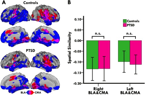
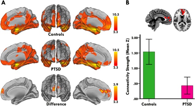
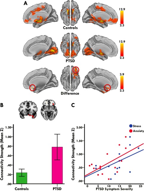
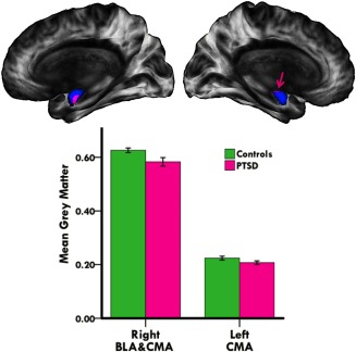
Posttraumatic stress disorder (PTSD) is a prevalent, debilitating, and difficult to treat psychiatric disorder. Very little is known of how PTSD affects neuroplasticity in the developing adolescent brain. Whereas multiple lines of research implicate amygdala-centered network dysfunction in the pathophysiology of adult PTSD, no study has yet examined the functional architecture of amygdala subregional networks in adolescent PTSD. Using intrinsic functional connectivity analysis, we investigated functional connectivity of the basolateral (BLA) and centromedial (CMA) amygdala in 19 sexually abused adolescents with PTSD relative to 23 matched controls. Additionally, we examined whether altered amygdala subregional connectivity coincides with abnormal grey matter volume of the amygdaloid complex. Our analysis revealed abnormal amygdalar connectivity and morphology in adolescent PTSD patients. More specifically, PTSD patients showed diminished right BLA connectivity with a cluster including dorsal and ventral portions of the anterior cingulate and medial prefrontal cortices (p < 0.05, corrected). In contrast, PTSD patients showed increased left CMA connectivity with a cluster including the orbitofrontal and subcallosal cortices (p < 0.05, corrected). Critically, these connectivity changes coincided with diminished grey matter volume within BLA and CMA subnuclei (p < 0.05, corrected), with CMA connectivity shifts additionally relating to more severe symptoms of PTSD. These findings provide unique insights into how perturbations in major amygdalar circuits could hamper fear regulation and drive excessive acquisition and expression of fear in PTSD. As such, they represent an important step toward characterizing the neurocircuitry of adolescent PTSD, thereby informing the development of reliable biomarkers and potential therapeutic targets.

Keywords: PTSD; adolescents; amygdala; grey matter; intrinsic functional connectivity.


Figures

Figure 1
Figure 1
Dissociable connectivity profiles of BLA and CMA complexes. (A) Lateral and medial views of the BLA (blue) and CMA (red) target networks in controls and adolescent PTSD patients. Overlap between BLA and CMA networks is shown in purple. Images are in radiological convention (i.e., right is left and vice versa). (B) Bar graph showing spatial correlations between BLA and CMA networks in controls and patients. Positive values indicate similarities while negative values indicate differences between BLA and CMA networks. Group comparison of spatial similarities between BLA and CMA target networks revealed no significant group differences. [Color figure can be viewed in the online issue, which is available at http://wileyonlinelibrary.com.]
Figure 2
Figure 2
Abnormal right BLA connectivity in adolescent PTSD patients. (A) Medial and anterior views of right BLA connectivity in controls (first row) and adolescent PTSD patients (second row). Direct group comparison revealed that PTSD patients had diminished right BLA connectivity with a cluster including dorsal and ventral portions of the anterior cingulate and medial prefrontal cortices (third row). (B) Representative sagittal and axial slices of between group differences in right BLA connectivity, along with a bar graph showing lower mean connectivity strength (i.e., mean Z) in PTSD patients within the displayed cluster. Statistical maps are corrected for multiple comparisons at the cluster level (Z > 2.3, p < 0.05), and brain images are in radiological convention (i.e., right is left and vice versa). [Color figure can be viewed in the online issue, which is available at http://wileyonlinelibrary.com.]
Figure 3
Figure 3
Abnormal left CMA connectivity in adolescent PTSD patients. (A) Medial and ventral views of left CMA connectivity in controls (first row) and adolescent PTSD patients (second row). Direct group comparison revealed that PTSD patients had increased left CMA connectivity with a cluster including the orbitofrontal and subcallosal cortices (third row). Red circles mark the effect site. (B) Representative coronal and axial slices of between group differences in left CMA connectivity, along with a bar graph showing higher mean connectivity strength (i.e., mean Z) in PTSD patients within the displayed cluster. Statistical maps are corrected for multiple comparisons at the cluster level (Z > 2.3, p < 0.05), and brain images are in radiological convention (i.e., right is left and vice versa). (C) Stronger CMA connectivity with the orbitofrontal/subcallosal region related to more symptoms of stress and anxiety in PTSD patients (r = 0.59 and 0.66; both p < 0.05). Individual patient's mean connectivity strength plotted against their symptom severity scores visualizes the direction of the association. PTSD‐related stress and anxiety was measured with the posttraumatic stress and anxiety subscales of the Trauma Symptom Checklist for Children (TSCC). [Color figure can be viewed in the online issue, which is available at http://wileyonlinelibrary.com.]
Figure 4
Figure 4
Abnormal grey matter volume of amygdala subregions in adolescent PTSD patients. Medial views of the right and left amygdaloid complex (blue), showing diminished grey matter volume of BLA (right hemisphere) and CMA (right and left hemispheres) subnuclei in adolescent PTSD patients (pink). Bar graph shows lower mean grey matter volume in PTSD patients within the displayed cluster. Right hemisphere results are corrected for multiple comparisons (p < 0.05, TFCE and FWE‐corrected), left hemisphere results are not (p < 0.05, TFCE‐corrected). Pink arrow marks the left CMA effect site. Brain images are in radiological convention (i.e., right is left and vice versa). [Color figure can be viewed in the online issue, which is available at http://wileyonlinelibrary.com.]

Similar articles

Cited by

References

    1. Ackerman PT, Newton JE, McPherson WB, Jones JG, Dykman RA (1998): Prevalence of post traumatic stress disorder and other psychiatric diagnoses in three groups of abused children (sexual, physical, and both). Child Abuse Negl 22:759–774. - PubMed
    1. Amunts K, Kedo O, Kindler M, Pieperhoff P, Mohlberg H, Shah NJ, Habel U, Schneider F, Zilles K (2005): Cytoarchitectonic mapping of the human amygdala, hippocampal region and entorhinal cortex: Intersubject variability and probability maps. Anat Embryol (Berl) 210:343–352. - PubMed
    1. Andreescu C, Sheu LK, Tudorascu D, Gross JJ, Walker S, Banihashemi L, Aizenstein H (2014): Emotion reactivity and regulation in late‐life generalized anxiety disorder: Functional connectivity at baseline and post‐treatment. Am J Geriatr Psychiatry - PMC - PubMed
    1. Armstrong JG, Putnam FW, Carlson EB, Libero DZ, Smith SR (1997): Development and validation of a measure of adolescent dissociation: The Adolescent Dissociative Experiences Scale. J Nerv Ment Dis 185:491–497. - PubMed
    1. Baas D, Aleman A, Kahn RS (2004): Lateralization of amygdala activation: A systematic review of functional neuroimaging studies. Brain Res Rev 45:96–103. - PubMed

Publication types

MeSH terms