. 2016 Jun;79(6):951-61.
doi: 10.1038/pr.2016.18. Epub 2016 Feb 9.

Smad7 interrupts TGF-β signaling in intestinal macrophages and promotes inflammatory activation of these cells during necrotizing enterocolitis

Affiliations

Smad7 interrupts TGF-β signaling in intestinal macrophages and promotes inflammatory activation of these cells during necrotizing enterocolitis

Krishnan MohanKumar et al. Pediatr Res. 2016 Jun.

Abstract

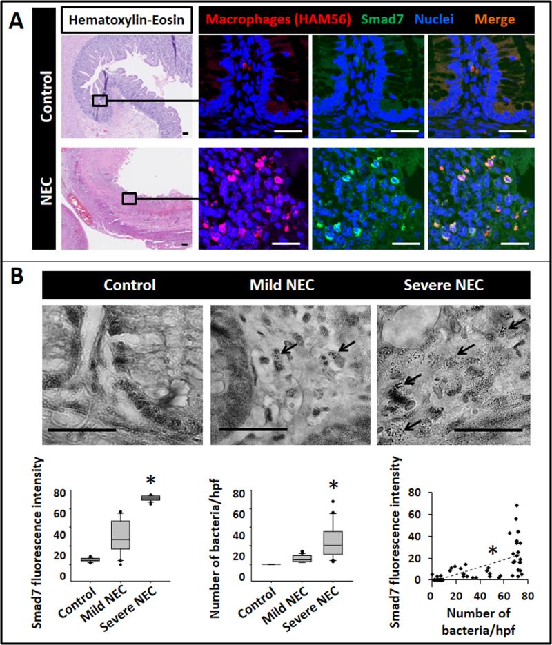
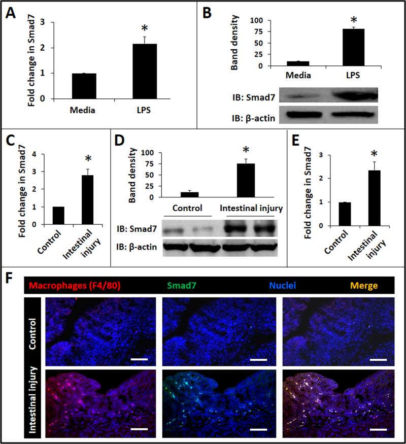
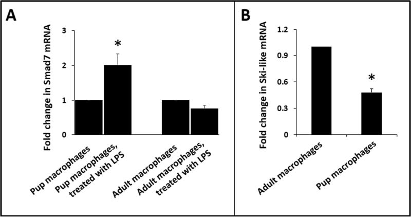
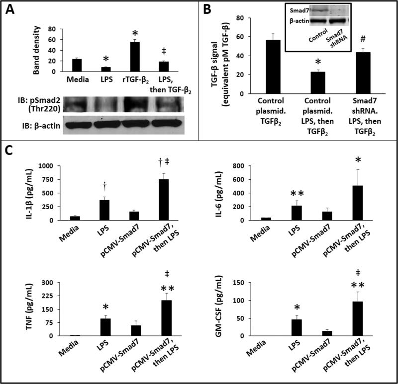
Background: Necrotizing enterocolitis (NEC) is an inflammatory bowel necrosis of premature infants. Based on our recent findings of increased Smad7 expression in surgically resected bowel affected by NEC, we hypothesized that NEC macrophages undergo inflammatory activation because increased Smad7 expression renders these cells resistant to normal, gut-specific, transforming growth factor (TGF)-β-mediated suppression of inflammatory pathways.

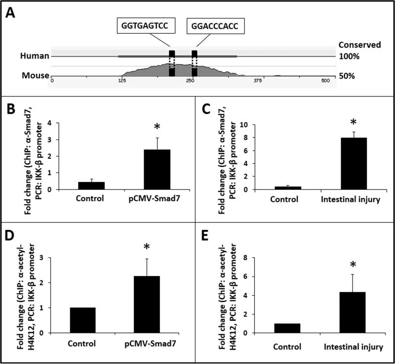
Methods: We used surgically resected human NEC tissue, murine models of NEC-like injury, bone marrow-derived and intestinal macrophages, and RAW264.7 cells. Smad7 and IκB kinase-beta (IKK-β) were measured by quantitative PCR, western blots, and immunohistochemistry. Promoter activation was confirmed in luciferase reporter and chromatin immunoprecipitation assays.

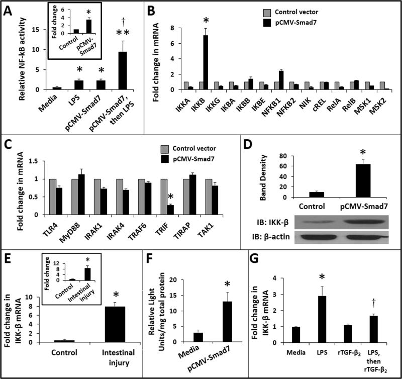
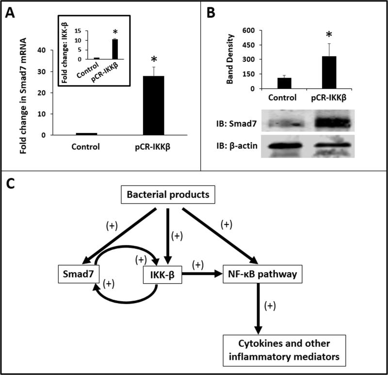
Results: NEC macrophages showed increased Smad7 expression, particularly in areas with severe tissue damage and high bacterial load. Lipopolysaccharide-induced Smad7 expression suppressed TGF-β signaling and augmented nuclear factor-kappa B (NF-κB) activation and cytokine production in macrophages. Smad7-mediated NF-κB activation was likely mediated via increased expression of IKK-β, which, further increased Smad7 expression in a feed-forward loop. We show that Smad7 induced IKK-β expression through direct binding to the IKK-β promoter and its transcriptional activation.

Conclusion: Smad7 expression in NEC macrophages interrupts TGF-β signaling and promotes NF-κB-mediated inflammatory signaling in these cells through increased expression of IKK-β.


Figures

Fig.1
Fig.1. Macrophages in NEC lesions express Smad7, particularly in areas with severe tissue damage and high bacterial load
(A) Top: Uninflamed premature intestine (ileum) showing intact crypt-villus histoarchitecture (hematoxylin & eosin, magnification 25×). Fluorescence photomicrographs on the right (650×) show macrophages (red; HAM56+) with no immunoreactivity for Smad7 (green). Bottom: NEC lesion with extensive necrosis and inflammation. Fluorescence photomicrographs on the right show macrophages with strong Smad7 immunoreactivity (green). Scale bar = 50 μm. Data represent 5 controls and 8 infants with NEC; (B) Representative photomicrographs (650×) show Brown and Brenn staining of uninflamed premature intestine, NEC with mild tissue damage, and NEC with severe tissue damage. Bacteria were seen in NEC lesions (arrows) but not in the uninflamed mucosa. Scale bar = 50 μm. N=8 infants with NEC; Box-whisker plots below show (left) increasing fluorescence intensity of Smad7 staining in macrophages with severity of tissue damage; (middle) greater bacterial load in areas with severe tissue injury than in regions with no or mild tissue damage; and (right) fluorescence intensity of Smad7 staining in macrophages increased with increasing number of bacteria per high-power field. Groups compared by Kruskal-Wallis H test; *P<0.01
Fig. 2
Fig. 2. Smad7 expression in LPS-treated RAW264.7 cells and in gut macrophages in NEC-like injury
(A) LPS increasedSmad7 expression in RAW264.7 macrophages. Bar-diagram (means ± SEM) shows fold change in Smad7 mRNA after treatment with 0.5 μg LPS/mL × 12h. Data represent 3 separate experiments; (B) Western blots confirm increased Smad7 expression in LPS-treated RAW264.7 cells at the protein level. Bar diagram (means ± SEM) summarizes densitometric data normalized against β-actin. Data represent 3 separate experiments; (C) Neonatal TNBSenterocolitis increased tissue expression of Smad7 mRNA. Bar diagram shows fold change in Smad7 mRNA in proximal colon from control vs. TNBS mice. N=5 animals/group; (D) Western blots confirmed increased Smad7 protein expression in TNBS colon. Bar diagram (means ± SEM) shows densitometric data normalized against β-actin. Data represent 3 separate experiments in different litters; (E) Increased Smad7 expression in macrophages isolated from ileocecal region of pups with TNBS-enterocolitis. Bar diagram (means ± SEM) show fold change in Smad7 mRNA. N=5 animals/group; (F) Fluorescence photomicrographs (100×) show localize Smad7 immunoreactivity in F4/80+ macrophages in TNBS-enterocolitis, but not in control. Scale bar = 50 μm. N=5 animals/group. Groups compared by Mann Whitney U test; * P<0.05.
Fig. 3
Fig. 3. Developmental differences in macrophage Smad7 expression
(A) LPS induced Smad7 in BMDMs from 10-day-old mouse pups but not in macrophages isolated from adult animals. Bar diagram (means ± SEM) shows fold change in Smad7 mRNA expression. (B) Macrophages from pups showed lower expression of Ski-like mRNA than adults. Bar diagram shows fold changes in mRNA. Data represent 3 separate experiments; Groups compared by the Kruskal-Wallis H test with pairwise comparisons; *P<0.05.
Fig. 4
Fig. 4. LPS-mediated suppression of TGF-β signaling in macrophages is mediated via Smad7
(A) LPS-treatment inhibited TGF-β-induced Smad2 phosphorylation in RAW264.7 cells. Western blots show phospho-Smad2 and β-actin expression in RAW264.7 macrophages treated with LPS (0.5 μg/mL overnight), TGF-β2 (500 pg/mL × 30 min), and LPS (0.5 μg/mL overnight) followed by TGF-β2 (500 pg/mL, 30 min). Bar diagram (means ± SEM) summarizes densitometric data from 3 separate experiments. *P<0.05 vs. media alone, ‡-P<0.05 vs. TGF-β2-treated cells; (B) Smad7 knockdown prior to LPS blocked LPS-mediated suppression of TGF-β signaling in macrophages. Bar diagram (means ± SEM) shows luciferase activity in SRE-reporter cells transfected with either control or Smad7-specific shRNA. These cells were treated overnight with LPS (or mock). Finally, we added rTGF-β2 × 1h and then measured TGF-β signaling. In the absence of LPS, Smad7 shRNA did not affect TGF-β signaling (not depicted). Data represent 3 separate experiments; Inset: Western blots confirmed Smad7 knockdown at protein level; (C) Smad7 overexpression increased LPS-induced cytokine expression in RAW264.7 cells. Bar diagrams (means ± SEM) show cytokine concentrations in supernatants: top left: IL-1β; top right: IL-6; bottom left: TNF; and bottom right: GM-CSF. Data represent 3 separate experiments. Groups compared by the Kruskal-Wallis H test with pairwise comparisons; * P<0.05, ** P<0.01 and †P<0.001 vs. media alone, ‡ P<0.05 vs. LPS-treated control cells.
Fig. 5
Fig. 5. Smad7 augments LPS-induced NF-κB activation in macrophages by inducing IKK-β expression in these cells
(A) Smad7 promoted LPS-mediated NF-κB activation in macrophages. Bar diagram (means ± SEM) shows relative NF-κB activity in control vs. Smad7-overexpressing NF-κB/SEAP reporter cells. Inset: Fold change (mean ± SEM) in Smad7 expression in macrophages following transfection with pCMV-Smad7; Data represent 3 experiments; * P<0.05, ** P<0.01 and †P<0.05 vs. pCMV-Smad7 transfected cells treated with media alone; (B) Smad7 overexpression in RAW264.7 macrophages induced IKK-β expression in these cells. Bar diagram (means ± SEM) shows fold change in mRNA expression of major NF-κB pathway genes. (C) Smad7-overexpression did not increase the expression of genes involved in TLR4-activated signaling; (D) Smad7 overexpression increased IKK-β protein expression in macrophages. Immunoblots show IKK-β expression in control and Smad7-overexpressing RAW264.7 cells. Bar diagram (means ± SEM) summarizes densitometric data, normalized against β-actin, from 3 experiments; (E) TNBS enterocolitis increased tissue expression of IKK-β. Bar diagram (means ± SEM) shows fold change in IKK-β mRNA in the proximal colon in TNBS-treated pups. N=5 pups per group. Inset: Increased IKK-β expression in intestinal macrophages isolated from the ileocecal region of TNBS pups; (F) Smad7 activated the IKK-β gene promoter. Bar diagram (means ± SEM) shows luciferase activity in control and Smad7-overexpressing RAW264.7 cells, transfected with a luciferase reporter carrying the IKK-β promoter. Data were normalized for total protein in the cell lysates; (G) rTGF-β2 suppression of LPS-induced IKK-β expression is not complete. Bar diagrams (means ± SEM) shows fold change in IKK-β mRNA in RAW264.7 cells in media alone, treated with LPS (0.5 μg/mL × 1h and then cultured overnight), rTGF-β2 (500 ng/mL, overnight), or LPS stimulation × 1h followed by rTGF-β2 overnight. * P<0.05, ** P<0.01 vs. control; †- P<0.05 vs. LPS-treated control cells. Two-group comparisons performed by the Mann-Whitney U test; Kruskal-Wallis H test (with pairwise comparisons) used for multiple groups.
Fig. 6
Fig. 6. Smad7 induces IKK-β expression in macrophages by directly activating its promoter
(A) In-silico analysis predicted 2 Smad-binding elements in human and murine IKK-β promoters; (B) Smad7 bound the IKK-β promoter in RAW264.7 macrophages. Bar diagram (means ± SEM) shows fold-enrichment of IKK-β promoter in LPS-treated native and Smad7-overexpressing RAW264.7 cells. Data represent 3 experiments; (C) Smad7 binding to IKK-β promoter was confirmed in tissue samples of TNBS-enterocolitis. N=5 animals/group; (D) Smad7 binding to the IKK-β promoter increased H4K12 acetylation on IKK-β nucleosome. Bar diagram (means ± SEM) show fold-enrichment of IKK-β promoter in ChIP using anti-acetyl-H4K12; (E) Increased H4K12 acetylation of the IKK-β promoter was confirmed in tissue samples of TNBS-enterocolitis. N=5 animals/group. Groups compared by the Mann-Whitney U test; * P<0.05.
Fig. 7
Fig. 7. IKK-β promotes Smad7 expression in macrophages and forms a positive feedback loop of inflammatory activation
(A) IKK-β overexpression induced Smad7 in macrophages. Bar diagram (means ± SEM) shows fold change in Smad7 mRNA in control vs. pCR-IKKβ-transfected RAW264.7 cells. Inset: Fold change (mean ± SEM) in IKK-β expression in macrophages transfected with pCR-IKKβ; (B) Immunoblots show Smad7 expression in LPS-treated control and IKKβ-overexpressing RAW264.7 cells. Bar diagram (means ± SEM) summarizes densitometric data, normalized against β-actin, from 3 experiments. Groups compared by the Mann-Whitney U test; * P<0.05. (C) Schematic showing the proposed signaling mechanisms for Smad7 effects on macrophages.

Similar articles

Cited by

References

    1. Neu J, Walker WA. Necrotizing enterocolitis. N Engl J Med. 2011;364:255–64. - PMC - PubMed
    1. Maheshwari A. Immunologic and Hematological Abnormalities in Necrotizing Enterocolitis. Clin Perinatol. 2015;42:567–85. - PMC - PubMed
    1. Remon JI, Amin SC, Mehendale SR, et al. Depth of bacterial invasion in resected intestinal tissue predicts mortality in surgical necrotizing enterocolitis. J Perinatol. 2015;35:755–62. - PMC - PubMed
    1. MohanKumar K, Kaza N, Jagadeeswaran R, et al. Gut mucosal injury in neonates is marked by macrophage infiltration in contrast to pleomorphic infiltrates in adult: evidence from an animal model. Am J Physiol Gastrointest Liver Physiol. 2012;303:G93–102. - PMC - PubMed
    1. Ballance WA, Dahms BB, Shenker N, Kliegman RM. Pathology of neonatal necrotizing enterocolitis: a ten-year experience. J Pediatr. 1990;117:S6–13. - PubMed