

Inhibition of CDK9 prevents mechanical injury-induced inflammation, apoptosis and matrix degradation in cartilage explants
- PMID: 26859911
- PMCID: PMC4750484
- DOI: 10.22203/ecm.v030a14
Inhibition of CDK9 prevents mechanical injury-induced inflammation, apoptosis and matrix degradation in cartilage explants
Abstract
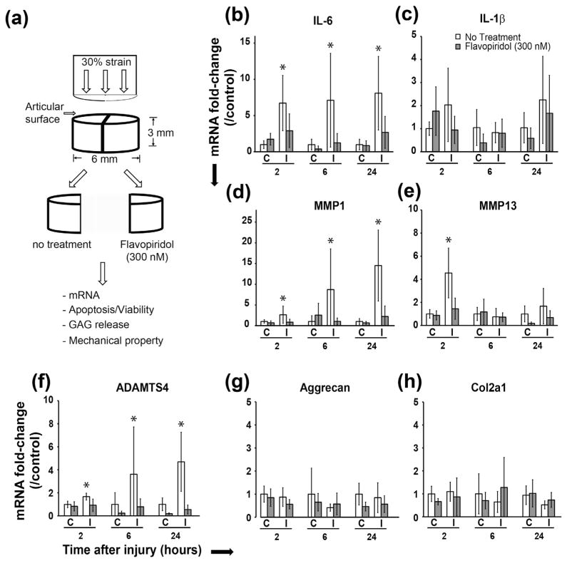
Joint injury often leads to post-traumatic osteoarthritis (PTOA). Acute injury responses to trauma induce production of pro-inflammatory cytokines and catabolic enzymes, which promote chondrocyte apoptosis and degrade cartilage to potentiate PTOA development. Recent studies show that the rate-limiting step for transcriptional activation of injury response genes is controlled by cyclin-dependent kinase 9 (CDK9), and thus it is an attractive target for limiting the injury response. Here, we determined the effects of CDK9 inhibition in suppressing the injury response in mechanically-injured cartilage explants. Bovine cartilage explants were injured by a single compressive load of 30 % strain at 100 %/s, and then treated with the CDK9 inhibitor Flavopiridol. To assess acute injury responses, we measured the mRNA expression of pro-inflammatory cytokines, catabolic enzymes, and apoptotic genes by RT-PCR, and chondrocyte viability and apoptosis by TUNEL staining. For long-term outcome, cartilage matrix degradation was assessed by soluble glycosaminoglycan release, and by determining the mechanical properties with instantaneous and relaxation moduli. Our data showed CDK9 inhibitor markedly reduced injury-induced inflammatory cytokine and catabolic gene expression. CDK9 inhibitor also attenuated chondrocyte apoptosis and reduced cartilage matrix degradation. Lastly, the mechanical properties of the injured explants were preserved by CDK9 inhibitor. Our results provide a temporal profile connecting the chain of events from mechanical impact, acute injury responses, to the subsequent induction of chondrocyte apoptosis and cartilage matrix deterioration. Thus, CDK9 is a potential disease-modifying agent for injury response after knee trauma to prevent or delay PTOA development.
Conflict of interest statement
The authors declare that they have no competing interest.
Figures


Similar articles
-
Bromodomain-containing-protein-4 and cyclin-dependent-kinase-9 inhibitors interact synergistically in vitro and combined treatment reduces post-traumatic osteoarthritis severity in mice.Osteoarthritis Cartilage. 2021 Jan;29(1):68-77. doi: 10.1016/j.joca.2020.07.012. Epub 2020 Oct 23. Osteoarthritis Cartilage. 2021. PMID: 33164842 Free PMC article.
-
Cyclin-dependent kinase 9 inhibition protects cartilage from the catabolic effects of proinflammatory cytokines.Arthritis Rheumatol. 2014 Jun;66(6):1537-46. doi: 10.1002/art.38378. Arthritis Rheumatol. 2014. PMID: 24470357 Free PMC article.
-
Effects of short-term glucocorticoid treatment on changes in cartilage matrix degradation and chondrocyte gene expression induced by mechanical injury and inflammatory cytokines.Arthritis Res Ther. 2011;13(5):R142. doi: 10.1186/ar3456. Epub 2011 Sep 2. Arthritis Res Ther. 2011. PMID: 21888631 Free PMC article.
-
Effects of shear stress on articular chondrocyte metabolism.Biorheology. 2000;37(1-2):95-107. Biorheology. 2000. PMID: 10912182 Review.
-
Impact of Abnormal Mechanical Stress on Chondrocyte Death in Osteoarthritis.Med Sci Monit. 2025 Jun 10;31:e948290. doi: 10.12659/MSM.948290. Med Sci Monit. 2025. PMID: 40493524 Free PMC article. Review.
Cited by
-
Cbx4 SUMOylates BRD4 to regulate the expression of inflammatory cytokines in post-traumatic osteoarthritis.Exp Mol Med. 2024 Oct;56(10):2184-2201. doi: 10.1038/s12276-024-01315-x. Epub 2024 Oct 1. Exp Mol Med. 2024. PMID: 39349832 Free PMC article.
-
Effects of TLR-2/NF-κB signaling pathway on the occurrence of degenerative knee osteoarthritis: an in vivo and in vitro study.Oncotarget. 2017 Jun 13;8(24):38602-38617. doi: 10.18632/oncotarget.16199. Oncotarget. 2017. PMID: 28418842 Free PMC article.
-
Bromodomain-containing-protein-4 and cyclin-dependent-kinase-9 inhibitors interact synergistically in vitro and combined treatment reduces post-traumatic osteoarthritis severity in mice.Osteoarthritis Cartilage. 2021 Jan;29(1):68-77. doi: 10.1016/j.joca.2020.07.012. Epub 2020 Oct 23. Osteoarthritis Cartilage. 2021. PMID: 33164842 Free PMC article.
-
Production of Inhalable Ultra-Small Particles for Delivery of Anti-Inflammation Medicine via a Table-Top Microdevice.Micromachines (Basel). 2022 Aug 25;13(9):1382. doi: 10.3390/mi13091382. Micromachines (Basel). 2022. PMID: 36144005 Free PMC article.
-
Intra-articular injection of flavopiridol-loaded microparticles for treatment of post-traumatic osteoarthritis.Acta Biomater. 2022 Sep 1;149:347-358. doi: 10.1016/j.actbio.2022.06.042. Epub 2022 Jun 30. Acta Biomater. 2022. PMID: 35779774 Free PMC article.
References
-
- Aigner T, Fundel K, Saas J, Gebhard PM, Haag J, Weiss T, Zien A, Obermayr F, Zimmer R, Bartnik E. Large-scale gene expression profiling reveals major pathogenetic pathways of cartilage degeneration in osteoarthritis. Arthritis Rheum. 2006;54:3533–3544. - PubMed
-
- Allen KD, Athanasiou KA. Viscoelastic characterization of the porcine temporomandibular joint disc under unconfined compression. J Biomech. 2006;39:312–322. - PubMed
-
- Amaral JD, Xavier JM, Steer CJ, Rodrigues CM. The role of p53 in apoptosis. Discov Med. 2010;9:145–152. - PubMed
-
- Bau B, Haag J, Schmid E, Kaiser M, Gebhard PM, Aigner T. Bone morphogenetic protein-mediating receptor-associated Smads as well as common Smad are expressed in human articular chondrocytes but not up-regulated or down-regulated in osteoarthritic cartilage. J Bone Miner Res. 2002;17:2141–2150. - PubMed
Publication types
MeSH terms
Substances
Grants and funding
LinkOut - more resources
Full Text Sources
Other Literature Sources
Miscellaneous