. 2016 Mar 2;8(8):5608-17.
doi: 10.1021/acsami.5b12400. Epub 2016 Feb 19.

Nanotubes-Embedded Indocyanine Green-Hyaluronic Acid Nanoparticles for Photoacoustic-Imaging-Guided Phototherapy

Affiliations

Nanotubes-Embedded Indocyanine Green-Hyaluronic Acid Nanoparticles for Photoacoustic-Imaging-Guided Phototherapy

Guohao Wang et al. ACS Appl Mater Interfaces. .

Abstract

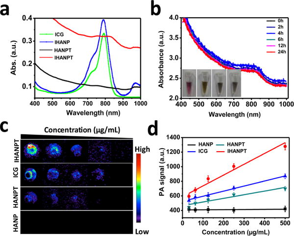
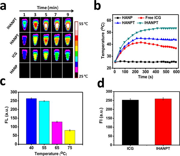
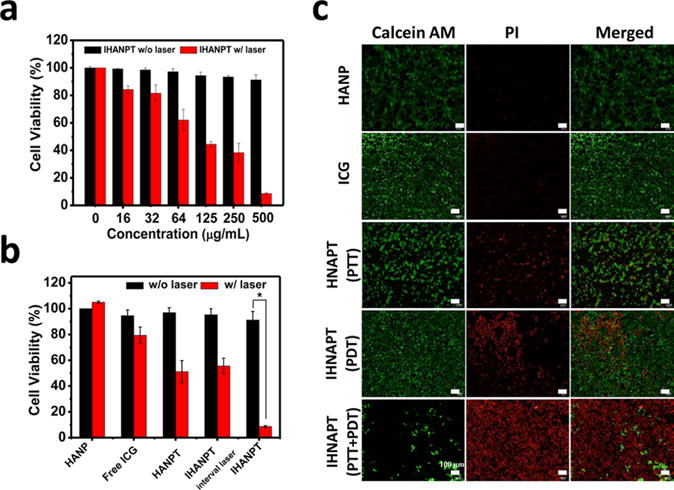
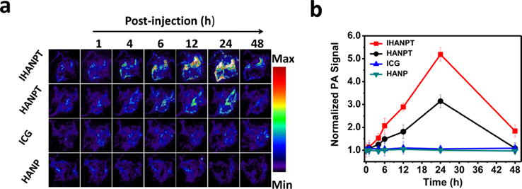
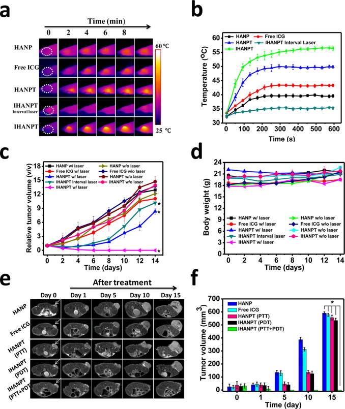
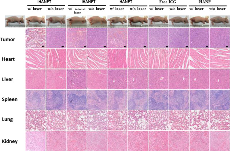
Phototherapy is a light-triggered treatment for tumor ablation and growth inhibition via photodynamic therapy (PDT) and photothermal therapy (PTT). Despite extensive studies in this area, a major challenge is the lack of selective and effective phototherapy agents that can specifically accumulate in tumors to reach a therapeutic concentration. Although recent attempts have produced photosensitizers complexed with photothermal nanomaterials, the tedious preparation steps and poor tumor efficiency of therapy still hampers the broad utilization of these nanocarriers. Herein, we developed a CD44 targeted photoacoustic (PA) nanophototherapy agent by conjugating Indocyanine Green (ICG) to hyaluronic acid nanoparticles (HANPs) encapsulated with single-walled carbon nanotubes (SWCNTs), resulting in a theranostic nanocomplex of ICG-HANP/SWCNTs (IHANPT). We fully characterized its physical features as well as PA imaging and photothermal and photodynamic therapy properties in vitro and in vivo. Systemic delivery of IHANPT theranostic nanoparticles led to the accumulation of the targeted nanoparticles in tumors in a human cancer xenograft model in nude mice. PA imaging confirmed targeted delivery of the IHANPT nanoparticles into tumors (T/M ratio = 5.19 ± 0.3). The effect of phototherapy was demonstrated by low-power laser irradiation (808 nm, 0.8 W/cm(2)) to induce efficient photodynamic effect from ICG dye. The photothermal effect from the ICG and SWCNTs rapidly raised the tumor temperature to 55.4 ± 1.8 °C. As the result, significant tumor growth inhibition and marked induction of tumor cell death and necrosis were observed in the tumors in the tumors. There were no apparent systemic and local toxic effects found in the mice. The dynamic thermal stability of IHANPT was studied to ensure that PTT does not affect ICG-dependent PDT in phototherapy. Therefore, our results highlight imaging property and therapeutic effect of the novel IHANPT theranostic nanoparticle for CD44 targeted and PA image-guided dual PTT and PDT cancer therapy.

Keywords: hyaluronic acid; indocyanine green; photodynamic therapy; photothermal therapy; single wall carbon nanotube.


Conflict of interest statement

Notes

The authors declare no competing financial interest.

Figures

Figure 1
Figure 1
Design and function of the dual targeted phototherapy agent, Indocyanine Green (ICG)-coupled threadlike nanoparticles (IHANPT).
Figure 2
Figure 2
Characterization of IHNAPT: (a) UV-vis-NIR absorbance spectra of free ICG, IHANP, HANPT, and IHANPT; (b) UV-vis absorbance spectra of IHANPT in darkness at 25 °C (inset shows the stability of IHANPT in water, PBS, fetal bovine serum (FBS), and cell medium after 2 weeks); (c) PAT images of IHNAPT, HNAPT, free ICG, and HANP at gradient diluted concentrations; and (d) standard curves of PAT signals in IHNAPT, HNAPT, free ICG, and HANP solution at different concentrations.
Figure 3
Figure 3
In vitro photothermal and 1O2 generation. (a) Thermal imaging profile of IHANPT, HANPT, ICG, and HANP, as a function of the irradiation time under continuous laser irradiation at a power intensity of 0.3 W/cm2. (b) Maximum temperature profiles of IHANPT, HANPT, ICG, and HANP, as a function of the irradiation time under continuous laser irradiation at a power intensity of 0.3 W/cm2. (c) 1O2 generation of IHANPT at different temperatures. (d) 1O2 generation of IHANP and ICG during single NIR laser irradiation (808 nm laser, light dose rate: 0.3 W/cm2).
Figure 4
Figure 4
Cytotoxicity: (a) cell viability studies with IHNAPT at different concentration with or without laser irradiation (808 nm, 0.3 W/cm2) on SCC7 cells; (b) cell viability studies with IHANPT (500 μg/mL IHANPT containing 70 μg/mL of SWCNTs, 16 μg/mL of ICG), HANPT, free ICG, and HANP with laser irradiation (808 nm, 0.3 W/cm2) on SCC7 cells (asterisk (*) denotes P < 0.05); (c) Calcein AM/PI staining to visualize SCC7 cell viability treated by with IHANPT, HANPT, free ICG with laser irradiation (808 nm, 0.3 W/cm2 for 10 min). Calcein AM (ex/em = 490/515 nm) staining of live cells is shown in green, and propidium iodide (PI) (ex/em = 535/615 nm) staining of dead cells is shown in red.
Figure 5
Figure 5
PA imaging: (a) in vivo PAT imaging of blood vessels in the tumor sites were captured at different time points after intravenous injection of IHANPT, HANPT, free ICG, and HANP; (b) photoacoustic intensity of tumor tissues at different time points.
Figure 6
Figure 6
In vivo phototherapy. (a) Thermal images of SCC7 tumor-bearing mice treated with HANP, free ICG, HANPT, and IHANPT (IHANPT containing 10 μg of SWCNTs and 5 μg of ICG, 100 μL) with 808 nm laser illumination at indicated time points. The laser power density was 0.8 W/cm2. (b) Quantitative analysis of temperature changes in tumor area at different time points. (c) Tumor growth curves of different groups of SCC7 tumor-bearing mice. Error bars represent the standard deviations of three mice per group. (Asterisk (*) denotes P < 0.05.) (d) Body weights were measured during the 14 day evaluation period in mice under different conditions. Dates indicate means and standard errors. (e) Magnetic resonance imaging (MRI)-monitored therapy response by IHANPT, IHANP, and ICG with laser irradiation. Arrows indicate the locations of tumors. (f) Tumor volume of different groups of SCC7 tumor-bearing mice measured by T2WI MRI after treatments. Error bars represent the standard deviations of three mice per group. (Asterisk (*) denotes P < 0.05.)
Figure 7
Figure 7
Histology staining. Hematoxylin and eosin (H&E) staining of tumors and primary organs of SCC7 tumor-bearing mice treated with IHANPT, HANPT, free ICG, and HANP with or without 808 nm NIR irradiation. Scale bars = 50 μm.

References

    1. Huang P, Lin J, Li W, Rong P, Wang Z, Wang S, Wang X, Sun X, Aronova M, Niu G, Leapman RD, Nie Z, Chen X. Biodegradable Gold Nanovesicles with an Ultrastrong Plasmonic Coupling Effect for Photoacoustic Imaging and Photothermal Therapy. Angew Chem. 2013;125:14208–14214. - PMC - PubMed
    1. Menon JU, Jadeja P, Tambe P, Vu K, Yuan B, Nguyen KT. Nanomaterials for Photo-Based Diagnostic and Therapeutic Applications. Theranostics. 2013;3:152–166. - PMC - PubMed
    1. Rai P, Mallidi S, Zheng X, Rahmanzadeh R, Mir Y, Elrington S, Khurshid A, Hasan T. Development and Applications of Photo-Triggered Theranostic Agents. Adv Drug Delivery Rev. 2010;62:1094–1124. - PMC - PubMed
    1. Zhang Z, Wang J, Chen C. Near Infrared Light Mediated Nanoplatforms for Cancer Thermo Chemotherapy and Optical Imaging. Adv Mater. 2013;25:3869–3880. - PubMed
    1. Zha Z, Wang J, Qu E, Zhang S, Jin Y, Wang S, Dai Z. Polypyrrole Hollow Microspheres as Echogenic Photothermal Agent for Ultrasound Imaging Guided Tumor Ablation. Sci Rep. 2013;3:2360. - PMC - PubMed

Publication types