

Contextual fear conditioning depresses infralimbic excitability
- PMID: 26860438
- PMCID: PMC4818676
- DOI: 10.1016/j.nlm.2016.01.015
Contextual fear conditioning depresses infralimbic excitability
Abstract
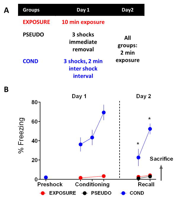
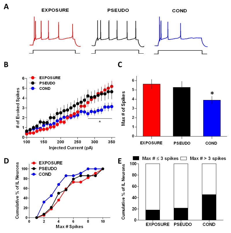
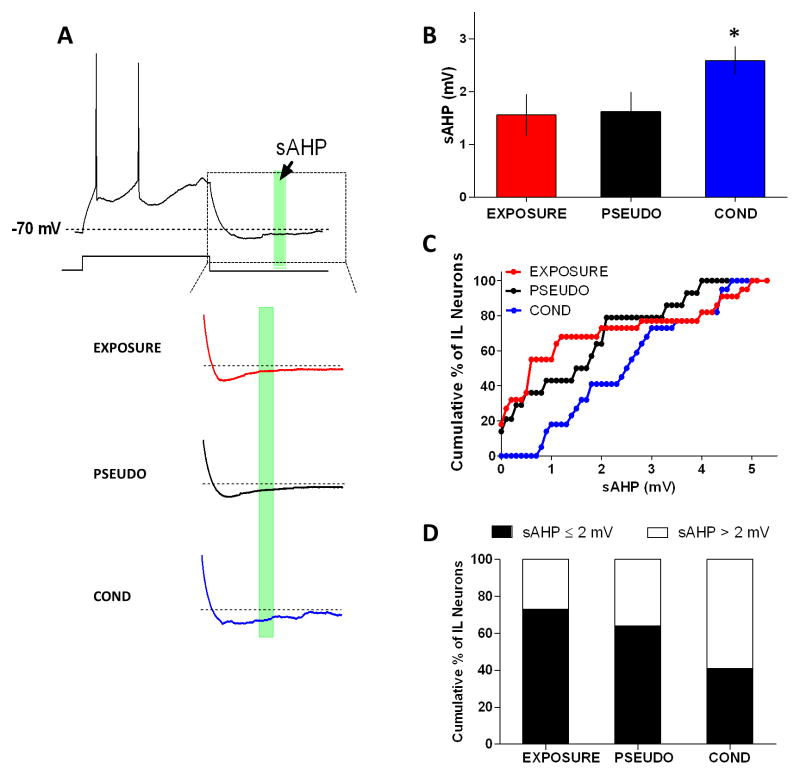
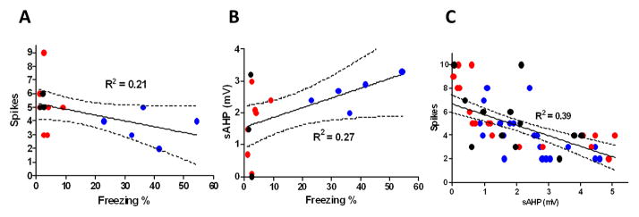
Patients with posttraumatic stress disorder (PTSD) show hypo-active ventromedial prefrontal cortices (vmPFC) that correlate with their impaired ability to discriminate between safe and dangerous contexts and cues. Previously, we found that auditory fear conditioning depresses the excitability of neurons populating the homologous structure in rodents, the infralimbic cortex (IL). However, it is undetermined if IL depression was mediated by the cued or contextual information. The objective of this study was to examine whether contextual information was sufficient to depress IL neuronal excitability. After exposing rats to context-alone, pseudoconditioning, or contextual fear conditioning, we used whole-cell current-clamp recordings to examine the excitability of IL neurons in prefrontal brain slices. We found that contextual fear conditioning reduced IL neuronal firing in response to depolarizing current steps. In addition, neurons from contextual fear conditioned animals showed increased slow afterhyperpolarization potentials (sAHPs). Moreover, the observed changes in IL excitability correlated with contextual fear expression, suggesting that IL depression may contribute to the encoding of contextual fear.
Keywords: Afterhyperpolarization; Brain slices; Contextual fear conditioning; Electrophysiology; Intrinsic excitability; Prefrontal cortex.
Copyright © 2016 Elsevier Inc. All rights reserved.
Figures
Similar articles
-
Fear conditioning and extinction differentially modify the intrinsic excitability of infralimbic neurons.J Neurosci. 2008 Apr 9;28(15):4028-36. doi: 10.1523/JNEUROSCI.2623-07.2008. J Neurosci. 2008. PMID: 18400902 Free PMC article.
-
Trace Fear Conditioning Differentially Modulates Intrinsic Excitability of Medial Prefrontal Cortex-Basolateral Complex of Amygdala Projection Neurons in Infralimbic and Prelimbic Cortices.J Neurosci. 2015 Sep 30;35(39):13511-24. doi: 10.1523/JNEUROSCI.2329-15.2015. J Neurosci. 2015. PMID: 26424895 Free PMC article.
-
M-type potassium channels modulate the intrinsic excitability of infralimbic neurons and regulate fear expression and extinction.J Neurosci. 2010 Sep 15;30(37):12379-86. doi: 10.1523/JNEUROSCI.1295-10.2010. J Neurosci. 2010. PMID: 20844133 Free PMC article.
-
Bidirectional changes in the intrinsic excitability of infralimbic neurons reflect a possible regulatory role in the acquisition and extinction of Pavlovian conditioned fear.J Neurosci. 2008 Jul 16;28(29):7245-7. doi: 10.1523/JNEUROSCI.2130-08.2008. J Neurosci. 2008. PMID: 18632927 Free PMC article. Review. No abstract available.
-
Learning-induced intrinsic and synaptic plasticity in the rodent medial prefrontal cortex.Neurobiol Learn Mem. 2020 Mar;169:107117. doi: 10.1016/j.nlm.2019.107117. Epub 2019 Nov 23. Neurobiol Learn Mem. 2020. PMID: 31765801 Free PMC article. Review.
Cited by
-
The Three Musketeers in the Medial Prefrontal Cortex: Subregion-specific Structural and Functional Plasticity Underlying Fear Memory Stages.Exp Neurobiol. 2022 Aug 31;31(4):221-231. doi: 10.5607/en22012. Exp Neurobiol. 2022. PMID: 36050222 Free PMC article. Review.
-
Infralimbic cortex controls fear memory generalization and susceptibility to extinction during consolidation.Sci Rep. 2020 Sep 28;10(1):15827. doi: 10.1038/s41598-020-72856-0. Sci Rep. 2020. PMID: 32985565 Free PMC article.
-
Sex differences in contextual fear expression are associated with altered medial prefrontal cortex activity.bioRxiv [Preprint]. 2024 Sep 13:2024.09.07.611834. doi: 10.1101/2024.09.07.611834. bioRxiv. 2024. PMID: 39314297 Free PMC article. Preprint.
-
Dynamic Changes of the Infralimbic Cortex and Its Regulation of the Prelimbic Cortex in Rats with Chronic Inflammatory Pain.Neurosci Bull. 2024 Jul;40(7):872-886. doi: 10.1007/s12264-023-01159-x. Epub 2024 Jan 5. Neurosci Bull. 2024. PMID: 38180711 Free PMC article.
-
Conditioning and pseudoconditioning differently change intrinsic excitability of inhibitory interneurons in the neocortex.Cereb Cortex. 2024 Apr 1;34(4):bhae109. doi: 10.1093/cercor/bhae109. Cereb Cortex. 2024. PMID: 38572735 Free PMC article.
References
-
- Beck H, Yaari Y. Plasticity of intrinsic neuronal properties in CNS disorders. Nat Rev Neurosci. 2008;9:357–369. - PubMed
-
- Bouton ME. Context and behavioral processes in extinction. Learn Mem. 2004;11:485–494. - PubMed
-
- Bouton ME, Bolles RC. Contextual control of the extinction of conditioned fear. Learn Motiv. 1979;10:445–466.
Publication types
MeSH terms
Grants and funding
LinkOut - more resources
Full Text Sources
Other Literature Sources
