. 2016 Mar 27;428(6):1180-1196.
doi: 10.1016/j.jmb.2016.01.031. Epub 2016 Feb 6.

Mapping the Interaction Network of Key Proteins Involved in Histone mRNA Generation: A Hydrogen/Deuterium Exchange Study

Affiliations

Mapping the Interaction Network of Key Proteins Involved in Histone mRNA Generation: A Hydrogen/Deuterium Exchange Study

Aleksandra Skrajna et al. J Mol Biol. .

Abstract

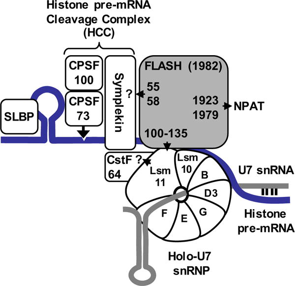
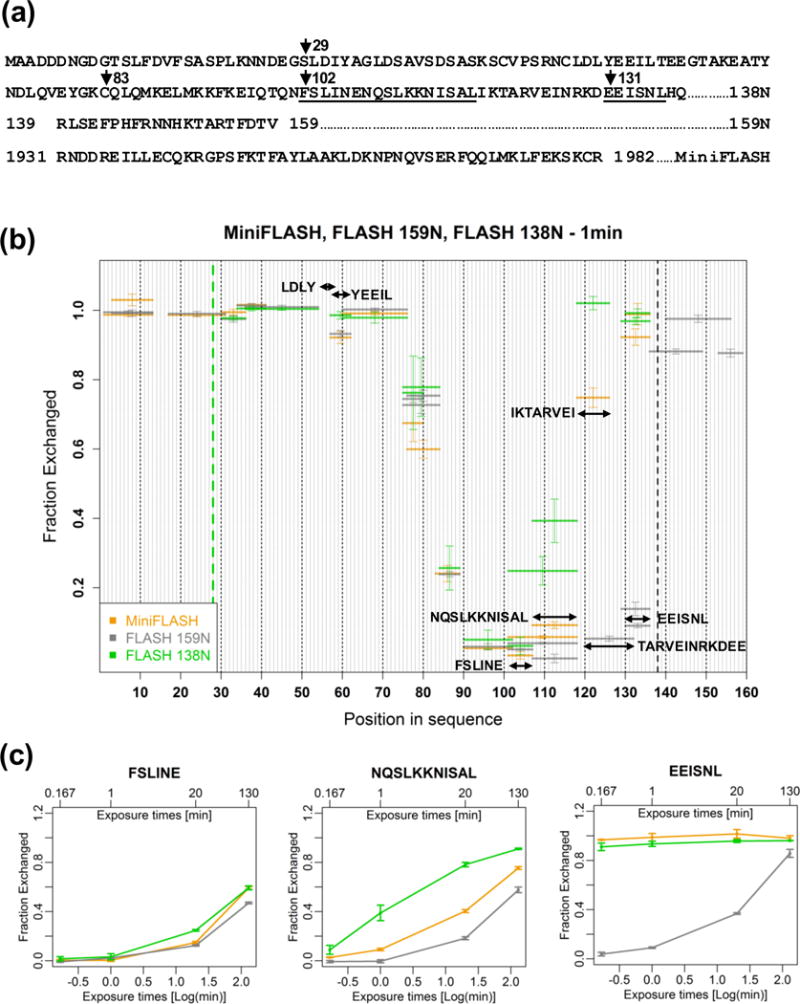
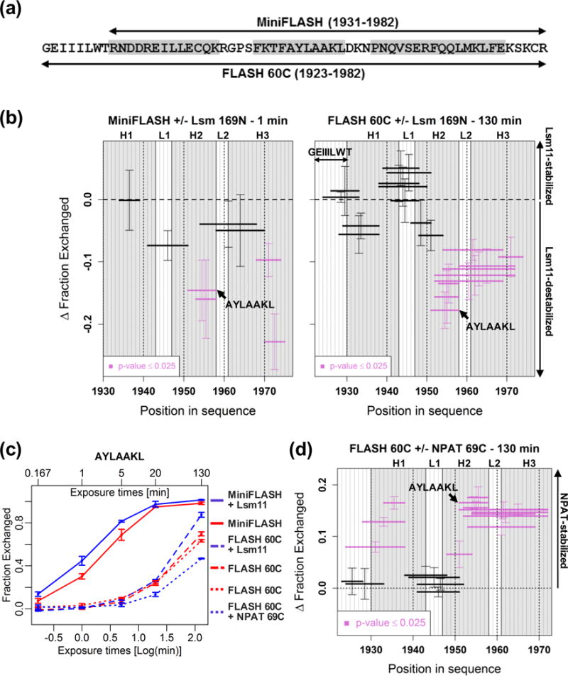
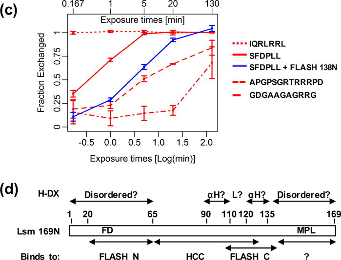
Histone pre-mRNAs are cleaved at the 3' end by a complex that contains U7 snRNP, the FLICE-associated huge protein (FLASH) and histone pre-mRNA cleavage complex (HCC) consisting of several polyadenylation factors. Within the complex, the N terminus of FLASH interacts with the N terminus of the U7 snRNP protein Lsm11, and together they recruit the HCC. FLASH through its distant C terminus independently interacts with the C-terminal SANT/Myb-like domain of nuclear protein, ataxia-telangiectasia locus (NPAT), a transcriptional co-activator required for expression of histone genes in S phase. To gain structural information on these interactions, we used mass spectrometry to monitor hydrogen/deuterium exchange in various regions of FLASH, Lsm11 and NPAT alone or in the presence of their respective binding partners. Our results indicate that the FLASH-interacting domain in Lsm11 is highly dynamic, while the more downstream region required for recruiting the HCC exchanges deuterium slowly and likely folds into a stable structure. In FLASH, a stable structure is adopted by the domain that interacts with Lsm11 and this domain is further stabilized by binding Lsm11. Notably, both hydrogen/deuterium exchange experiments and in vitro binding assays demonstrate that Lsm11, in addition to interacting with the N-terminal region of FLASH, also contacts the C-terminal SANT/Myb-like domain of FLASH, the same region that binds NPAT. However, while NPAT stabilizes this domain, Lsm11 causes its partial relaxation. These competing reactions may play a role in regulating histone gene expression in vivo.

Keywords: FLASH; Lsm11; Mass spectrometry; NPAT; U7 snRNP.


Figures

Fig. 1
Fig. 1. Key proteins involved in histone gene expression
Known components of U7-dependent complex that cleaves histone pre-mRNA (blue line) at the 3′ end. 5′ end of U7 snRNA (grey line) base pairs with histone pre-mRNA. The U7 snRNA and the heptameric Sm ring together form the core U7 snRNP that upon interacting with FLASH recruits the HCC, giving rise to holo-U7 snRNP. Only four essential subunits of the HCC are shown: symplekin, CPSF100, CPSF73 and CstF64. Numbers in FLASH indicate amino acids that are required for the interaction of FLASH with the HCC, Lsm11 and NPAT. The arrangement of the four polyadenylation subunits in the HCC is hypothetical. The identity of the subunits that interact with FLASH and Lsm11 has not been determined, as indicated by question marks.
Fig. 2
Fig. 2. Hydrogen/Deuterium (H/D) exchange in the N-terminal region of FLASH
A. Sequences of the three FLASH variants, each containing a single GST tag at the N-terminus. FLASH 138N has amino acids 1–28 deleted. With the exception of cysteine 83 being mutated to alanine in FLASH 138N and FLASH 159N, the three proteins are identical in the region 29–138. In MiniFLASH, the first 138 amino acids are fused to the C-terminal region of FLASH (amino acids 1931-1982) as a result of alternative splicing. Peptides analyzed in panel C are underlined. B. H/D exchange patterns for FLASH 138N (green), FLASH 159N (grey) and MiniFLASH (orange) after 1 min incubation in D2O buffer. The horizontal lines indicate individual peptides and the error bars are standard deviations from at least three independent experiments. The green and black dashed lines delimit the N- and C-terminal regions, respectively, that differ among the three FLASH variants. The C-terminal domain of MiniFLASH is not shown in the plot. C. Time-dependent H/D exchange for three selected peptides (102FSLINE107, 108NQSLKKNISAL118 and 131EEISNL136) from FLASH 138N (green), FLASH 159N (grey) and MiniFLASH (orange). H/D exchange was analyzed at four time points plotted on a logarithmic scale. Time in minutes (0.167 min, 1 min, 20 min, 130 min) is shown at the top of the graph. Error bars represent standard deviations calculated from at least three independent experiments.
Fig. 3
Fig. 3. H/D exchange in FLASH in the apo-state and bound to Lsm11
A. FLASH 159N and an equimolar mixture of FLASH 159N and Lsm11 (amino acids 1–169) were incubated in D2O for 130 min and the extent of H/D exchange for the same peptides in the unbound (red) and Lsm11-bound (blue) state was determined by mass spectrometry. Results shown for each peptide are an average of at least three independent experiments, with the error bars representing standard deviations. The 108NQSLKKNISALIK120 peptide is indicated with an arrow and amino acid 138 is indicated with a black dashed line. B. Differential plot of H/D exchange presented in panel A. Δ Fraction Exchanged was calculated by subtracting Fraction Exchanged values for FLASH 159N in the presence of Lsm 169N (bound state, blue) from the values obtained in the apo-state (red). Peptides with p-values below 0.025 in the t-test are labeled in bright purple.
Fig. 4
Fig. 4. Different effects of Lsm11 and NPAT binding on the SANT/Myb-like domain
A. Amino acid sequence of the C-terminal region of FLASH in MiniFLASH (top) and FLASH 60C (bottom). Predicted α-helical structures are shown in grey. B. Differential plots of H/D exchange within the C-terminal domain of FLASH in MiniFLASH (left panel) and FLASH 60C (right panel). Δ Fraction Exchanged was calculated as described in Fig. 3B. α-helical structures (H1, H2 and H3) are marked in grey. Loop regions are indicated with L1 and L2. Positive and negative values indicate stabilization and destabilization of the C-terminal FLASH region, respectively. C. Kinetic plots of H/D exchange for the 1952AYLAAKL1958 peptide in MiniFLASH and FLASH 60C, either in the apo-state (red) or in a complex with Lsm 169N or NPAT 69C (blue), as indicated. D. Differential plots of H/D exchange for FLASH 60C bound to equimolar amounts of NPAT 69C after 130 min incubation in D2O buffer.
Fig. 5
Fig. 5. Binding of Lsm11 and NPAT to the C-terminal region of FLASH is mutually-exclusive
A. BLASTP-generated amino acid sequence alignment of the C-terminal regions of human FLASH and YARP. Non-identical amino acids that are unlikely to affect the overall structure of each protein are indicated with the plus sign. α-helices are underlined. B. GST-tagged proteins indicated at the top of each lane were incubated with 35S-labeled Lsm11 169N (top panels) or NPAT 131C (bottom panels) and their binding analyzed by the GST pull down assay. Proteins were collected on glutathione beads, separated on a SDS/polyacrylamide gel and detected by either staining with Coomassie blue (GST proteins, bottom panels) or by autoradiography (35S-labeled proteins, top panels). C. Binding of GST-tagged YARP 97C and FLASH 60C, either alone (lanes 2 and 4) or pre-bound to NPAT 69C (lanes 3 and 5), to 35S-labeled Lsm11 169N.
Fig. 6
Fig. 6. Mapping the region in Lsm11 that interacts with the C-terminal FLASH domain
A. Amino acid sequence of the N-terminal region of human Lsm11 (1–169). Black arrows indicate end points of various deletion mutants used in the pull down experiments. The FD dipeptide critical for the interaction of Lsm11 with the N-terminal FLASH and the highly stable GDGAAGAGRRG peptide (see Fig. 7) are underlined. B. Binding of GST-tagged FLASH 138N or FLASH 60C (lanes 2 and 3, respectively) to 35S-labeled Lsm11 169N or its FD-AA mutant. C. Binding of GST-tagged FLASH 138N or FLASH 60C (lanes 2 and 3, respectively) to 35S-labeled Lsm11 169N (top panels) or its deletion mutants: 151N (middle panels) or 142N (bottom panels). D. Binding of GST-tagged FLASH 138N, FLASH 60C or YARP 97C to 35S-labeled Lsm11 105N.
Fig. 7
Fig. 7. H/D exchange in Lsm11 bound to FLASH
A. H/D exchange for Lsm11 (amino acids 1-169) in the apo-state (red) and bound to an equimolar amount of FLASH 138N (blue) after 1 min incubation in D2O buffer. Peptides analyzed in panel C are indicated with the arrows. B. Differential plot of H/D exchange presented in panel A. C. Time-dependent H/D exchange for indicated Lsm11 peptides. The 30SFDPLL35 peptide is shown in both the apo-state (red) and in a complex with FLASH 138N (blue). D. Localization of potential functional and structural elements within the N-terminal region of Lsm11 (amino acids 1-169), as predicted based on the H/D exchange and GST-pull down assays.
Fig. 8
Fig. 8. A possible set of events that occur at the G1/S phase transition, resulting in activation of histone gene expression
FLASH and NPAT interact through their C-terminal regions (indicated by the double-headed arrow), forming a repressive complex that prevents each protein from performing its function in histone gene expression. Phosphorylation of NPAT by Cyclin E/CDK2 at the end of the G1 phase disrupts this complex and activates both transcription of histone genes by liberating NPAT and 3′ end processing of histone transcripts by promoting FLASH-dependent assembly of the active U7 snRNP. The Lsm11-binding sites in the N- and C-terminal region of FLASH are indicated with a long and short vertical arrow, respectively.

Similar articles

Cited by

References

    1. Marzluff WF. Metazoan replication-dependent histone mRNAs: a distinct set of RNA polymerase II transcripts. Curr Opin Cell Biol. 2005;17:274–280. - PubMed
    1. Ma TL, Van Tine BA, Wei Y, Garrett MD, Nelson D, Adams PD, Wang J, Qin J, Chow LT, Harper JW. Cell cycle-regulated phosphorylation of p220NPAT by cyclin E/Cdk2 in Cajal bodies promotes histone gene transcription. Genes and Development. 2000;14:2298–2313. - PMC - PubMed
    1. Ye X, Wei Y, Nalepa G, Harper JW. The cyclin E/Cdk2 substrate p220(NPAT) is required for S-phase entry, histone gene expression, and Cajal body maintenance in human somatic cells. Mol Cell Biol. 2003;23:8586–8600. - PMC - PubMed
    1. Zhao JY, Kennedy BK, Lawrence BD, Barbie DA, Matera AG, Fletcher JA, Harlow E. NPAT links cyclin E-Cdk2 to the regulation of replication-dependent histone gene transcription. Genes and Development. 2000;14:2283–2297. - PMC - PubMed
    1. Miele A, Braastad CD, Holmes WF, Mitra P, Medina R, Xie R, Zaidi SK, Ye X, Wei Y, Harper JW, Van Wijnen AJ, Stein JL, Stein GS. HiNF-P directly links the cyclin E/CDK2/p220NPAT pathway to histone H4 gene regulation at the G1/S phase cell cycle transition. Mol Cell Biol. 2005;25:6140–6153. - PMC - PubMed

Publication types

LinkOut - more resources