

Coexistence of splenic hemangioma and vascular malformation of the vertebrae
- PMID: 26860733
- PMCID: PMC4748470
- DOI: 10.1186/s13104-016-1860-6
Coexistence of splenic hemangioma and vascular malformation of the vertebrae
Abstract
Background: Cavernous hemangioma is an encapsulated mass of dilated, endothelial lined vascular channels filled with slowly flowing blood. Cavernous hemangioma of the spleen is a rare condition with less than 100 reports so far. Hemangioma of the vertebral is a benign vascular legion around one or two vertebrae. These are usually asymptomatic and discovered incidentally. In this study we reported an extreme rare case of splenic hemangioma coexistence with vascular malformation of the vertebrae. To our knowledge this is the first report of coexistence of splenic hemangioma and hemangioma of the vertebra.
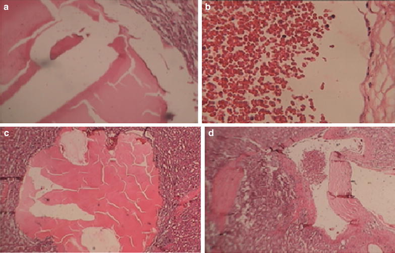
Case presentation: A 20-year-old iranian male with splenomegaly, abdominal pain, diarrhea and pancytopenia who was first highly suspicious for malignancy referred to our center for evaluation of the diagnostic workup. After full examination we detected a very rare case with a giant, solitary cavernous hemangioma of the spleen and multiple hemangiomas in his vertebrae. Histopathology of the spleen showed a large cavernous hemangioma occupying almost the entire spleen with large areas of infarction necrosis with multiple hemangiomas of the vertebrae.
Conclusion: It is extremely rare to have a splenic hemangioma concurrent with vertebra hemangioma and this is clinically very important to consider splenic hemangioma in differential diagnosis of splenomegaly for a better therapeutic management in related patients.
Figures


References
-
- Abbott RM, Levy AD, Aguilera NS, Gorospe L, Thompson WM. From the archives of the AFIP: primary vascular neoplasms of the spleen: radiologic–pathologic correlation. Radiographics. 2004;24(1137–11):63. - PubMed
-
- Zinner M, Ashley JS. Maingot’s Abdominal operations, 11th ed.NewYork: Mcgraw hill; 2007.
Publication types
MeSH terms
LinkOut - more resources
Full Text Sources
Other Literature Sources
Medical
