

Deletion of Men1 and somatostatin induces hypergastrinemia and gastric carcinoids
- PMID: 26860771
- PMCID: PMC4980289
- DOI: 10.1136/gutjnl-2015-310928
Deletion of Men1 and somatostatin induces hypergastrinemia and gastric carcinoids
Erratum in
-
Erratum: Deletion of Men1 and somatostatin induces hypergastrinemia and gastric carcinoids.Gut. 2017 Nov;66(11):2012. doi: 10.1136/gutjnl-2015-310928corr1. Gut. 2017. PMID: 28993466 No abstract available.
Abstract
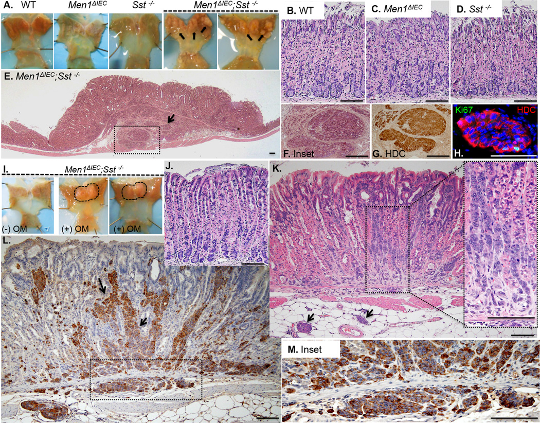
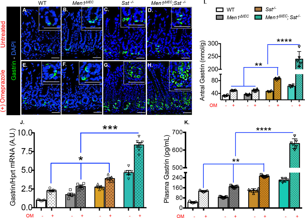
Background: Gastric carcinoids are slow growing neuroendocrine tumours arising from enterochromaffin-like (ECL) cells in the corpus of stomach. Although most of these tumours arise in the setting of gastric atrophy and hypergastrinemia, it is not understood what genetic background predisposes development of these ECL derived tumours. Moreover, diffuse microcarcinoids in the mucosa can lead to a field effect and limit successful endoscopic removal.
Objective: To define the genetic background that creates a permissive environment for gastric carcinoids using transgenic mouse lines.
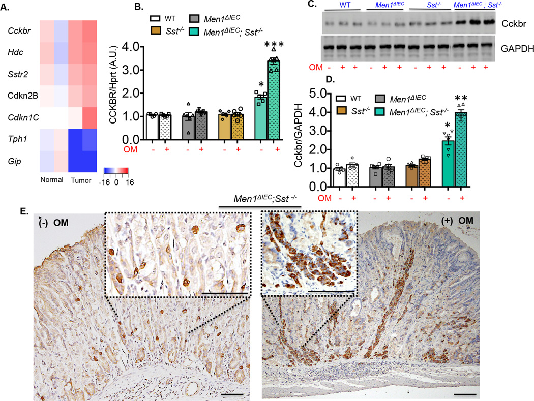
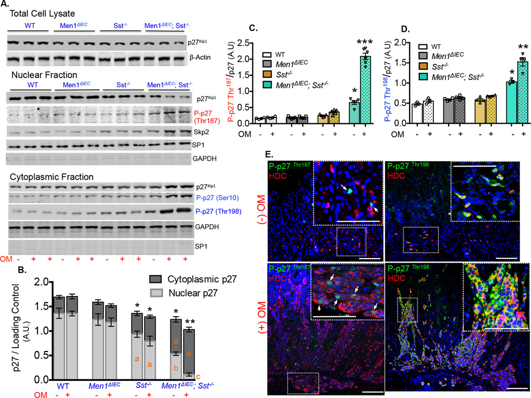
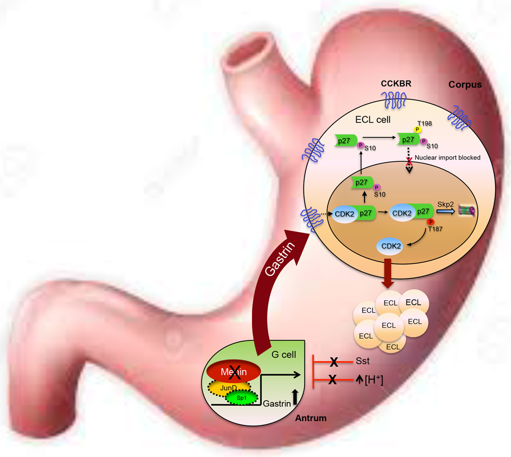
Design: The multiple endocrine neoplasia 1 gene locus (Men1) was deleted using Cre recombinase expressed from the Villin promoter (Villin-Cre) and was placed on a somatostatin null genetic background. These transgenic mice received omeprazole-laced chow for 6 months. The direct effect of gastrin and the gastrin receptor antagonist YM022 on expression and phosphorylation of the cyclin inhibitor p27Kip1 was tested on the human human gastric adenocarcinoma cell line stably expressing CCKBR (AGSE) and mouse small intestinal neuroendocrine carcinoma (STC)-1 cell lines.
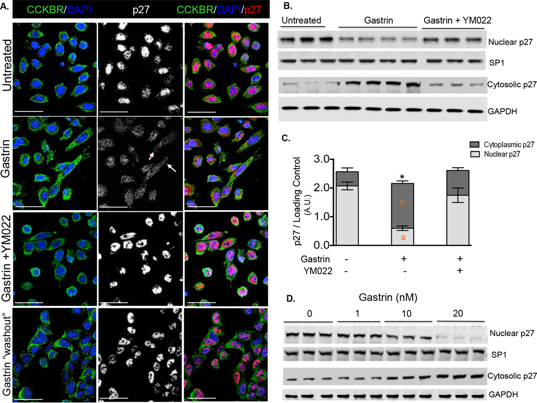
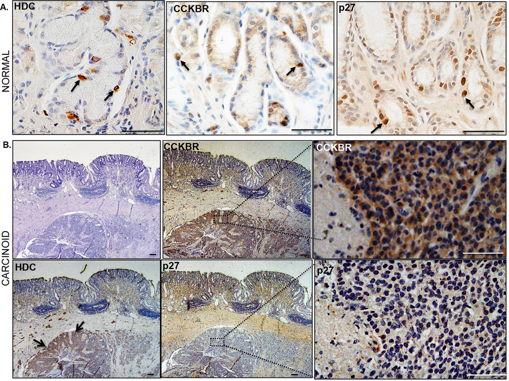
Results: The combination of conditional Men1 deletion in the absence of somatostatin led to the development of gastric carcinoids within 2 years. Suppression of acid secretion by omeprazole accelerated the timeline of carcinoid development to 6 months in the absence of significant parietal cell atrophy. Carcinoids were associated with hypergastrinemia, and correlated with increased Cckbr expression and nuclear export of p27Kip1 both in vivo and in gastrin-treated cell lines. Loss of p27Kip1 was also observed in human gastric carcinoids arising in the setting of atrophic gastritis.
Conclusions: Gastric carcinoids require threshold levels of hypergastrinemia, which modulates p27Kip1 cellular location and stability.
Keywords: ACID SECRETION; CANCER GENETICS; ENDOCRINE TUMOURS; GASTRIN; GASTRIN RECEPTORS.
Published by the BMJ Publishing Group Limited. For permission to use (where not already granted under a licence) please go to http://www.bmj.com/company/products-services/rights-and-licensing/.
Figures
Similar articles
-
Gastrin Induces Nuclear Export and Proteasome Degradation of Menin in Enteric Glial Cells.Gastroenterology. 2017 Dec;153(6):1555-1567.e15. doi: 10.1053/j.gastro.2017.08.038. Epub 2017 Aug 30. Gastroenterology. 2017. PMID: 28859856 Free PMC article.
-
Altered influence of CCK-B/gastrin receptors on HDC expression in ECL cells after neoplastic transformation.Regul Pept. 1999 Dec 23;85(2-3):115-23. doi: 10.1016/s0167-0115(99)00086-5. Regul Pept. 1999. PMID: 10651065
-
Pathophysiology of Gastric NETs: Role of Gastrin and Menin.Curr Gastroenterol Rep. 2017 Jul;19(7):32. doi: 10.1007/s11894-017-0572-y. Curr Gastroenterol Rep. 2017. PMID: 28608155 Free PMC article. Review.
-
Hypergastrinemia and gastric enterochromaffin-like cells.Am J Surg Pathol. 1995;19 Suppl 1:S8-19. Am J Surg Pathol. 1995. PMID: 7762739 Review.
-
Netazepide Inhibits Expression of Pappalysin 2 in Type 1 Gastric Neuroendocrine Tumors.Cell Mol Gastroenterol Hepatol. 2020;10(1):113-132. doi: 10.1016/j.jcmgh.2020.01.010. Epub 2020 Jan 28. Cell Mol Gastroenterol Hepatol. 2020. PMID: 32004755 Free PMC article. Clinical Trial.
Cited by
-
Preclinical Models of Neuroendocrine Neoplasia.Cancers (Basel). 2022 Nov 17;14(22):5646. doi: 10.3390/cancers14225646. Cancers (Basel). 2022. PMID: 36428741 Free PMC article. Review.
-
Uncovering the role of traditional Chinese medicine in immune-metabolic balance of gastritis from the perspective of Cold and Hot: Jin Hong Tablets as a case study.Chin Med. 2024 Oct 4;19(1):134. doi: 10.1186/s13020-024-00998-8. Chin Med. 2024. PMID: 39367502 Free PMC article.
-
Gastrin Induces Nuclear Export and Proteasome Degradation of Menin in Enteric Glial Cells.Gastroenterology. 2017 Dec;153(6):1555-1567.e15. doi: 10.1053/j.gastro.2017.08.038. Epub 2017 Aug 30. Gastroenterology. 2017. PMID: 28859856 Free PMC article.
-
Low Pepsinogen I/II Ratio and High Gastrin-17 Levels Typify Chronic Atrophic Autoimmune Gastritis Patients With Gastric Neuroendocrine Tumors.Clin Transl Gastroenterol. 2020 Sep;11(9):e00238. doi: 10.14309/ctg.0000000000000238. Clin Transl Gastroenterol. 2020. PMID: 33094954 Free PMC article.
-
Weight gain in mice on a high caloric diet and chronically treated with omeprazole depends on sex and genetic background.Am J Physiol Gastrointest Liver Physiol. 2017 Jan 1;312(1):G15-G23. doi: 10.1152/ajpgi.00211.2016. Epub 2016 Nov 3. Am J Physiol Gastrointest Liver Physiol. 2017. PMID: 27810953 Free PMC article.
References
-
- Burkitt MD, Pritchard DM. Review article: Pathogenesis and management of gastric carcinoid tumours. Aliment Pharmacol Ther. 2006;24:1305–1320. - PubMed
-
- Gilligan CJ, Lawton GP, Tang LH, West AB, Modlin IM. Gastric carcinoid tumors: the biology and therapy of an enigmatic and controversial lesion. Am J Gastroenterol. 1995;90:338–352. - PubMed
-
- Meeker A, Heaphy C. Gastroenteropancreatic endocrine tumors. Mol Cell Endocrinol. 2014;386:101–120. - PubMed
-
- Bakke I, Qvigstad G, Sandvik AK, Waldum HL. The CCK-2 receptor is located on the ECL cell, but not on the parietal cell. Scand J Gastroenterol. 2001;36:1128–1133. - PubMed
-
- Norton JA, Melcher ML, Gibril F, Jensen RT. Gastric carcinoid tumors in multiple endocrine neoplasia-1 patients with Zollinger-Ellison syndrome can be symptomatic, demonstrate aggressive growth, and require surgical treatment. Surgery. 2004;136:1267–1274. - PubMed
Publication types
MeSH terms
Substances
Grants and funding
LinkOut - more resources
Full Text Sources
Other Literature Sources
Medical
Molecular Biology Databases
Miscellaneous