. 2016 May;44(5):352-7.
doi: 10.1016/j.exphem.2016.01.009. Epub 2016 Feb 6.

Leukemia and chromosomal instability in aged Fancc-/- mice

Affiliations

Leukemia and chromosomal instability in aged Fancc-/- mice

Donna Cerabona et al. Exp Hematol. 2016 May.

Abstract

Fanconi anemia (FA) is an inherited disorder of genomic instability associated with high risk of myelodysplasia and acute myeloid leukemia (AML). Young mice deficient in FA core complex genes do not naturally develop cancer, hampering preclinical studies on malignant hematopoiesis in FA. Here we describe that aging Fancc(-/-) mice are prone to genomically unstable AML and other hematologic neoplasms. We report that aneuploidy precedes malignant transformation during Fancc(-/-) hematopoiesis. Our observations reveal that Fancc(-/-) mice develop hematopoietic chromosomal instability followed by leukemia in an age-dependent manner, recapitulating the clinical phenotype of human FA and providing a proof of concept for future development of preclinical models of FA-associated leukemogenesis.


Figures

Fig 1
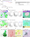
Fig 1. Aging Fancc−/− mice develop hematologic malignancies
(A) Kaplan-Meier survival curve of wt (n=20) and Fancc−/− (n=18) cohorts. (B) Table demonstrating incidence of leukemias and lymphomas in wt and Fancc−/− mice by 24 months of age. Statistical significance for (A, B) was determined using a log-rank (Mantel-Cox) test. Peripheral blood smear of a moribund Fancc−/− mouse (C) shows leukemic blasts (arrows). Diagnosis of acute myeloid leukemia was confirmed with flow cytometry demonstrating increased expression of the Gr1 myeloid marker in the peripheral blood compared to wt and healthy Fancc−/− controls (D) and the presence of leukemic infiltrates in the liver (E). Bone marrow cytospin (F) of another moribund Fancc−/− mouse demonstrated multiple blasts (arrows). Flow cytometry demonstrated increased expression of the B220 B-cell marker on bone marrow blasts (G) and necropsy revealed leukemic infiltrates in the liver (H), consistent with B-cell ALL. Necropsy of another Fancc−/− mouse demonstrated conglomerates of mesenteric lymph nodes (I, J). Liver infiltrates in this mouse were Cd3+ (K, L), consistent with T-cell lymphoma.
Fig 2
Fig 2. Transplanted Fancc−/− AML LDMNCs induce aggressive leukemia in wt recipients
(A) Competitive repopulation transplant schematic. Cd45.2+ LDMNCs obtained from a leukemic Fancc−/− mouse or wt control were mixed 1:1 with Cd45.1+ wt competitor LDMNCs and transplanted into lethally irradiated wt recipients (n=3 recipients per genotype). (B) Recipients of Fancc−/− LDMNCs died within 2 months after transplantation. Log-rank (Mantel-Cox) test was used to assess significance. (C) AML in wt recipients of Fancc−/− LDMNCs. Gr1 was used as a myeloid marker for flow cytometry (right panel). (D) Splenomegaly in moribund wt mice transplanted with Fancc−/− LDMNCs. (E, F) Leukemic Cd45.2+ Fancc−/− LDMNCs overpopulate recipient bone marrow compared to wt Cd45.1+ competitor LDMNCs. Statistical significance was determined using an unpaired student's t-test; error bars represent SEM.
Fig 3
Fig 3. Genomic instability and abnormal mitosis in leukemic and pre-leukemic Fancc−/− mice
(A) Representative images of LDMNC metaphase spreads from wt and leukemic Fancc−/− mice. (B) Increased aneuploidy in leukemic Fancc−/− LDMNCs. At least 74 spreads were counted from wt, Fancc−/− non-leukemic, and Fancc−/− leukemic mice. Note increased chromosomal instability in non-leukemic Fancc−/− LDMNCs compared to age-matched wt controls. Fisher's exact test was used to determine statistical significance. Leukemic Fancc−/− LDMNCs undergo abnormal mitosis (C) and have a higher mitotic index (D) compared to LDMNCs from wt and Fancc−/− non-leukemic mice (n=3 mice/genotype; at least 500 cells were counted per genotype). Statistical analyses were performed using chi-square tests with Yates correction.

References

    1. Kottemann MC, Smogorzewska A. Fanconi anaemia and the repair of Watson and Crick DNA crosslinks. Nature. 2013;493(7432):356–363. - PMC - PubMed
    1. Nalepa G, Clapp DW. Fanconi anemia and the cell cycle: new perspectives on aneuploidy. F1000Prime Rep. 2014;6:23. - PMC - PubMed
    1. D'Andrea AD. Susceptibility pathways in Fanconi's anemia and breast cancer. N Engl J Med. 2010;362(20):1909–1919. - PMC - PubMed
    1. Alter BP. Fanconi anemia and the development of leukemia. Best Pract Res Clin Haematol. 2014;27(3-4):214–221. - PMC - PubMed
    1. Parmar K, D'Andrea A, Niedernhofer LJ. Mouse models of Fanconi anemia. Mutat Res. 2009;668(1-2):133–140. - PMC - PubMed

Publication types

Substances