Review
. 2016 Feb 6;17(2):170.
doi: 10.3390/ijms17020170.

Focus on Extracellular Vesicles: Introducing the Next Small Big Thing

Affiliations
Review

Focus on Extracellular Vesicles: Introducing the Next Small Big Thing

Hina Kalra et al. Int J Mol Sci. .

Abstract

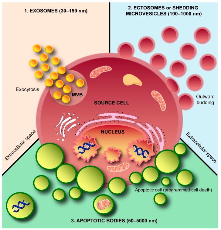
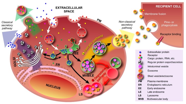
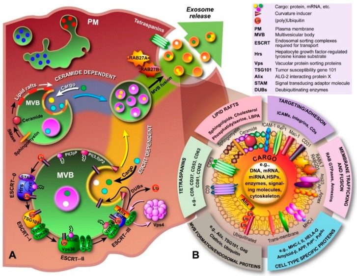
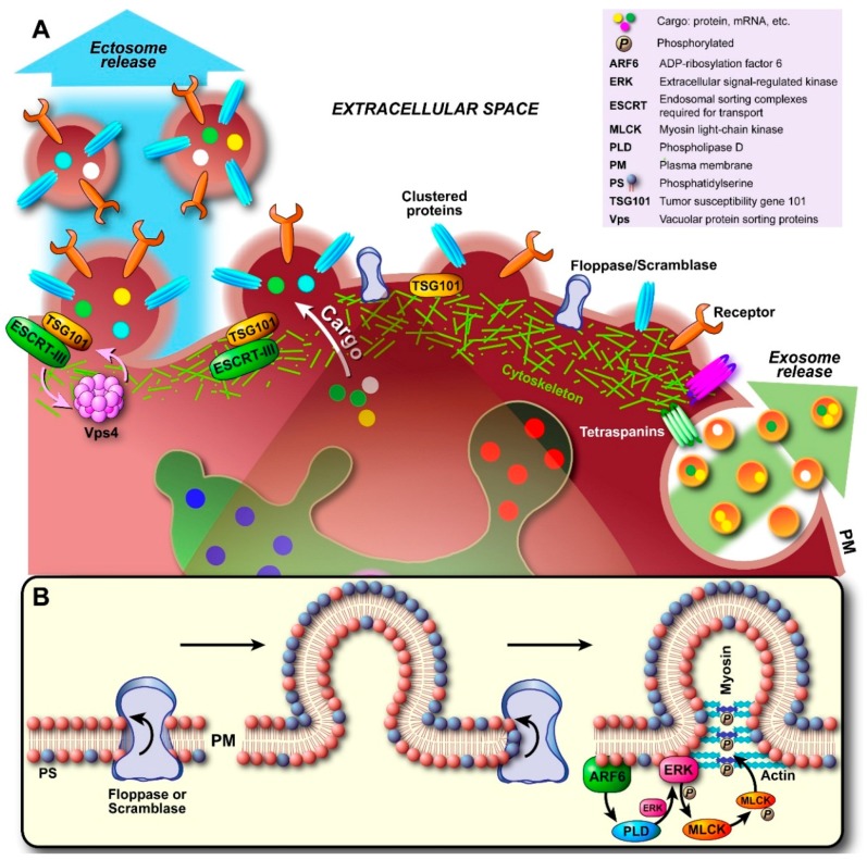
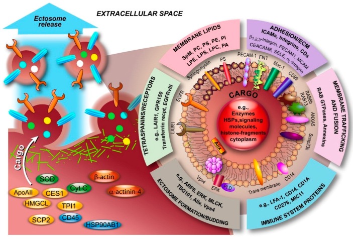
Intercellular communication was long thought to be regulated exclusively through direct contact between cells or via release of soluble molecules that transmit the signal by binding to a suitable receptor on the target cell, and/or via uptake into that cell. With the discovery of small secreted vesicular structures that contain complex cargo, both in their lumen and the lipid membrane that surrounds them, a new frontier of signal transduction was discovered. These "extracellular vesicles" (EV) were initially thought to be garbage bags through which the cell ejected its waste. Whilst this is a major function of one type of EV, i.e., apoptotic bodies, many EVs have intricate functions in intercellular communication and compound exchange; although their physiological roles are still ill-defined. Additionally, it is now becoming increasingly clear that EVs mediate disease progression and therefore studying EVs has ignited significant interests among researchers from various fields of life sciences. Consequently, the research effort into the pathogenic roles of EVs is significantly higher even though their protective roles are not well established. The "Focus on extracellular vesicles" series of reviews highlights the current state of the art regarding various topics in EV research, whilst this review serves as an introductory overview of EVs, their biogenesis and molecular composition.

Keywords: apoptotic body; biogenesis; ectosome; exosome; extracellular vesicle; isolation; microvesicle; molecular composition; signal transduction.


Figures

Figure 1
Figure 1
Schematic representation of subtypes of extracellular vesicles (EVs) released by a cell. Three subtypes of EVs, namely exosomes, shedding microvesicles or ectosomes and apoptotic bodies, are known to be secreted by a cell into the extracellular space. Exosomes are released by exocytosis, whereas shedding microvesicles or ectosomes are secreted by outward budding of the plasma membrane. Apoptotic bodies are released by dying cells during the later stages of apoptosis so that cell debris can easily be eliminated by neighboring and immune system cells. MVB: multivesicular body.
Figure 2
Figure 2
Pathways involving various types of vesicles. In the classical secretory pathway, vesicles with protein cargo, sorted and packed in the Golgi apparatus, transport their cargo to the plasma membrane (PM). By fusing with the PM, both membrane proteins and secretory proteins are effectively transported to their intended destinations. Various types of cargo, e.g., proteins, RNA, can also be transported into the extracellular space via outward PM budding and formation of shedded vesicles (ectosomes). Cargo is taken up by the cell via endocytosis (receptor-mediated and free uptake) and formation of early endosomes. In early endosomes, proteins are either recycled to the PM or sequestered into the intraluminal vesicles (ILV) of MVBs. Formation of exosomes starts with inward budding of the early endosome’s membrane and subsequent formation of MVBs. In the exocytic pathway ①, MVBs fuse with the PM to release their contents (exosomes) into the extracellular space; In the degradative pathway ②, the MVBs are trafficked to lysosomes for enzyme-assisted degradation. This pathway is particularly important for restricting signaling by activated growth factor receptors. Exosomal cargo delivery to the recipient cell can occur through various mechanisms, i.e., direct fusion with the recipient cell’s membrane, pinocytosis/phagocytosis, or ligand–receptor binding.
Figure 3
Figure 3
Biogenesis, secretion and composition of exosomes. (A) The biogenesis and secretion of exosomes is believed to be mediated via a ceramide and/or ESCRT-dependent pathway. The ceramide-dependent pathway is based on the formation of lipid rafts in which sphingomyelin is converted to ceramide by sphingomyelinases. These ceramide-enriched domains have structural imbalances between monoleaflets causing the membrane to bend inward. In the ESCRT-dependent pathway, components of the ESCRT machinery are sequentially recruited to the endosomal membrane, which starts with Hrs, and bind to phosphatidylinositol-3-phosphate (PI(3)P) and the 3,5-bisphosphate (PI(3,5)P2) through lipid binding domains (e.g., FYVE, GLUE), and to the ubiquitinated protein (ESCRT-0). ESCRT-I and -II drive budding of ILVs, during which cargo is transported into the lumen, and ESCRT-III is recruited by Alix to complete budding and drive vesicle scission (spiral formation and pulling). DUBs deubiquitinate the protein and Vps4 recycles the ESCRT machinery. The now formed MVB is transported to the PM and through fusion, the ILVs are released into the extracellular environment and are now called “exosomes”; (B) Exosomal luminal cargo predominantly consists of mRNA, miRNA and gDNA fragments, and a myriad of different proteins depending on the cell of origin. Generally, proteins involved in MVB formation, tetraspanins, membrane transport and fusion, transmembrane proteins, cytoskeletal components and proteins of cytosolic origin are part of exosomes. In addition, biomolecules associated with various diseases, including cancer, neurodegenerative diseases, such as Parkinson’s, Alzheimer’s and transmissible spongiform encephalopathies (prion disease), and inflammatory disorders have been identified in exosomes.
Figure 4
Figure 4
Biogenesis and secretion of ectosomes. (A) Initial nucleation at the plasma membrane (PM) starts with clustering of transmembrane proteins and lipids in distinct domains. Recruited and PM associated proteins such as tetraspanins (largely still unidentified) may be involved in sorting of components analogous to exosomal sorting. Additionally, Ca2+ release/accumulation and activation of enzymes induce degradation of cytoskeletal components. Outward budding is promoted by externalization of phosphatidylserine (PS) by specific translocases (floppase, scramblase; see also (B)). As the cytoskeleton disintegrates locally and becomes more traversable, cytosolic proteins and genetic material are sorted into the lumen. Budding and pinching off are generally thought to occur either through the model proposed in (B), where budding involves initiation of a signaling cascade by ARF6 through activation and recruitment of PLD/ERK and phosphorylation of MLCK. This triggers actomyosin contraction and pinching off of the ectosome. Alternatively, recent evidence suggests that recruited TSG101 induces translocation of ESCRT-III to the PM, which in turn results in conical spiral assembly (budding initiator), and finally Vps4 ATPase constriction of the ring of ESCRT-III spirals at the budding neck leads to membrane scission and pinching off, as shown in (A).
Figure 5
Figure 5
Molecular composition of ectosomes. Ectosomal membranes consist of various classes of lipids. Furthermore, in recent years, numerous components with diverse functions have been identified, predominantly from blood, immune and endothelial cells, and atherosclerotic plaques. The overview serves to illustrate this diversity and is far from complete. Data was retrieved from Vesiclepedia (Available online: http://www.microvesicles.org). ANXA5, annexin A5; ApoAII, apolipoprotein AII; ARF6, ADP-ribosylation factor 6; CD45, protein tyrosine phosphatase; CEACAM8, carcinoembryonic antigen-related cell adhesion molecule 8; CES1, carboxylesterase 1; Cyt C, cytochrome C; ECM, extracellular matrix; EGFRvIII, mutated form of epidermal growth factor receptor; ERK, extracellular-signal-regulated kinase; FN1, fibronectin 1; GPR150, G protein-coupled receptor 150; HMGCL, 3-hydroxymethyl-3-methylglutaryl-coenzyme A lyase; HSP90AB1, heat shock protein 90 kDa alpha (cytosolic) class B member 1; LAIR1, leukocyte-associated immunoglobulin-like receptor 1; LFA-1, lymphocyte function-associated antigen 1; LPC, lysohosphatidylcholine; LPE, lysophosphatidylethanolamine; LPS, lysophosphatidylserine; Mac-1, macrophage-1 antigen; MCAM, melanoma cell adhesion molecule; MLCK, myosin light-chain kinase; PA, phosphatidic acid; PC, phosphatidylcholine; PE, phosphatidylethanolamine; PECAM1, platelet/endothelial cell adhesion molecule 1; PI, phosphatidylinositol; PLD, phospholipase D; PS, phosphatidylserine; SCP2, sterol carrier protein 2; SELE, selectin E; Snap23b, synaptosomal-associated protein 23b; SOD, superoxide dismutase; SpM, sphingomyelin; TPI1, triosephosphate isomerase 1.

References

    1. Raposo G., Stoorvogel W. Extracellular vesicles: Exosomes, microvesicles, and friends. J. Cell Biol. 2013;200:373–383. doi: 10.1083/jcb.201211138. - DOI - PMC - PubMed
    1. Simons M., Raposo G. Exosomes-vesicular carriers for intercellular communication. Curr. Opin. Cell Biol. 2009;21:575–581. doi: 10.1016/j.ceb.2009.03.007. - DOI - PubMed
    1. Balaj L., Lessard R., Dai L., Cho Y.-J., Pomeroy S.L., Breakefield X.O., Skog J. Tumour microvesicles contain retrotransposon elements and amplified oncogene sequences. Nat. Commun. 2011;2 doi: 10.1038/ncomms1180. - DOI - PMC - PubMed
    1. Cossetti C., Iraci N., Mercer T.R., Leonardi T., Alpi E., Drago D., Alfaro-Cervello C., Saini H.K., Davis M.P., Schaeffer J. Extracellular vesicles from neural stem cells transfer IFN-γ via Ifngr1 to activate Stat1 signaling in target cells. Mol. Cell. 2014;56:193–204. doi: 10.1016/j.molcel.2014.08.020. - DOI - PMC - PubMed
    1. Théry C., Zitvogel L., Amigorena S. Exosomes: Composition, biogenesis and function. Nat. Rev. Immunol. 2002;2:569–579. - PubMed

Publication types